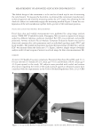
PRECLINICAL SURFACTANT SKIN PENETRATION ASSAY 221 an average molecular weight of 30,000 Da and 17% of unhydrolyzed acetate groups. Common commercial PVAs have an acetate content of 4–12% (9). Radiolabeled SDS (14C-SDS, 55 mCi/mmol) was obtained from American Radiolabeled Chemicals (St. Louis, MO). Tritiated water (3H2O, 1.0 mCi/ml) and the tissue-dissolution reagents Sol- uene®-350 and Solvable™ were obtained from Perkin Elmer (Waltham, MA). Dulbecco’s phosphate-buffered saline (PBS) and sodium azide were obtained from Fisher Scientifi c (Pittsburgh, PA). Deionized (DI) water was prepared by ultrafi ltration. D-Squame™ tapes were obtained from CuDerm (Dallas, TX). Pig skin was obtained from a local slaughter house and dermatomed to a thickness of ~800 μm. Human cadaver skin, der- matomed to a thickness of 300–400 μm, was obtained from the New York Firefi ghters Skin Bank (New York, NY). A different donor was used in each experimental trial. The source and identity of each human donor skin sample (age, ethnicity, gender, date of death, and cause of death) was documented. The pig skin studies were approved by the P&G Institutional Animal Care and Use Committee, and work on de-identifi ed human tissues was exempted from human subjects’ categorization by the University of Cincin- nati Academic Health Center Institutional Review Board. PREPARATION OF SKIN MEMBRANES Human skin was stored at -80°C until use. On the morning before the study, the skin was thawed rapidly by immersing the sealed packet in warm water. It was then rinsed with distilled water and cut into 2 × 2 cm pieces using a scalpel. Porcine skin taken from the belly area was obtained from a slaughterhouse, stored in chilled saline, and used within 24 h of collection. IN VITRO STATIC DIFFUSION CELLS The skin membranes were mounted in Franz diffusion cells (0.79 cm2) (10) with the SC facing the donor chamber. The receptor solution (~5 ml) was Dulbecco’s PBS (pH 7.4) to which 0.02% w:v sodium azide had been added to retard microbial growth. The receptor fl uid was continuously stirred using a magnetic stir bar. The cells were maintained at 37°C in a thermostatted aluminum block, yielding a skin-surface temperature of 32°C. Low glass tops with no occlusion were used for this study. HUMAN SKIN MEMBRANE INTEGRITY ASSESSMENT The integrity of each of the human skin membranes was assessed by 3 H2O penetration (8). The skin was mounted and allowed to equilibrate for about 1 h. A 150 μl aliquot of 3 H2O (0.4 μCi/ml) was applied using a pipette and allowed to remain on the skin surface for 5 min. It was then removed with a cotton-tipped swab, which was placed on the skin surface for 30 s. The receptor solution was collected 60 min after dose and replaced with PBS. The collected samples were analyzed for 3 H in Ultima Gold XR cocktail (Perkin Elmer, Waltham, MA) by liquid scintillation counting (LSC) using a Beckman LS 6500 counter (Beckman Coulter, Inc., Indianapolis, IN). They were counted for 1 min, and the results

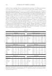
JOURNAL OF COSMETIC SCIENCE 222 were reported as μl 3 H2O/cm2. Samples with water permeation greater than 2.0 μl 3 H2O/ cm2 were discarded. The remaining cells were ranked in order of increasing water perme- ability to facilitate the random controlled block experimental design (8). The receptor exchange procedure was repeated, and the cells were allowed to wash out overnight. A fi nal exchange was performed in the morning before dosing. Porcine skin integrity was assessed visually to ensure the absence of large hair follicles. SURFACTANT PENETRATION PROTOCOL—PORCINE SKIN 14 C-SDS solutions (50 mM SLS + 6.7 μCi/ml 14 C-SDS) in DI water, with and without 2% w/w of added polymer, were prepared and shaken to ensure homogeneity. The SLS con- centration corresponds to 1.44% w/v, about 10-fold lower than typical anionic surfactant concentrations in a shampoo or shower gel. This is a commonly accepted dilution factor for consumer exposures. The test concentration was furthermore about 16-fold higher than the apparent CMC for the SLS sample, so most of the SLS in these formulations existed in either micellar or polymer-bound micellar form. A 150 μl aliquot (10 μCi) of the surfac- tant solution was pipetted onto each skin membrane. Skin from one donor was exposed to the surfactant solution for 10 min (n = 6/treatment). After the surfactant exposure, the dose solution was removed using a transfer pipet. The surface of the skin was rinsed three times with 0.5 ml of tap water for 10 s, and the rinses were collected and pooled. The receptor solution was collected, and each skin sample was wiped two times with Whatman fi lter paper (GE Healthcare Life Sciences, Pittsburgh, PA) soaked with PBS/Tween 20 and once with 70%/30% ethanol/water to remove unabsorbed (residual) product. Wipes were collected and pooled for mass bal- ance determination. After surface rinsing, the surfaces of the skins were dried, and 10 tape strips (D-Squame™) were collected. The tapes were placed directly into Ultima Gold XR cocktail to be analyzed individually. After tape stripping, the remaining epi- dermis was dissected from the dermis, and the skin sections were dissolved in 0.50–1.25 ml Soluene-350™ at 50°C overnight. Radioactivity in receptor collections, surface rinses, fi lter paper wipes, tape strips, and solubilized tissue sections was determined using LSC. Results were expressed as μg/cm2 14 C-SDS equivalents or % of applied radioactive dose. The arithmetic mean and standard error mean (SEM) were reported for each treatment. SURFACTANT PENETRATION PROTOCOL—HUMAN SKIN 14 C-SDS solutions (50 mM SLS + 6.7 μCi/ml 14 C-SDS) in DI water, with and without 2% w/w of an added polymer, were prepared and shaken to ensure homogeneity. A 150 μl aliquot (10 μCi) of the surfactant solution was pipetted onto each skin membrane, which were rank ordered in terms of permeability based on the 3 H2O prescreening results. The rankordering and subsequent randomization by treatment were key elements in maxi- mizing the sensitivity of the assay (8). Two sets of experiments were conducted. In Experiment 1, skin from three donors was exposed to the surfactant solution for 10 min (n = 4–6/donor). The total sample size was n = 14–15/treatment. In Experiment 2, skin from four donors was exposed to the surfac-
Purchased for the exclusive use of nofirst nolast (unknown)
From: SCC Media Library & Resource Center (library.scconline.org)