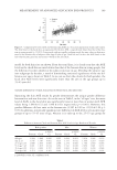
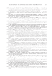
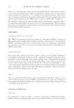
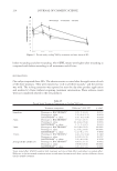
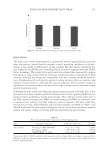
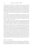
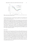
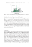
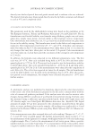
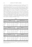
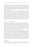
PRECLINICAL SURFACTANT SKIN PENETRATION ASSAY 229 slowly than surfactant alone their deposition onto the SC surface in a consumer-relevant 2-min exposure would thus be reduced relative to surfactant alone. The alternative scenario of micellar penetration through pores in the SC presented by Moore et al. (1) and supported by other articles from this group (3,7) is not ruled out by the previous argument. But, it seems to us that it is not necessary to invoke the presence of microscopic pores of a specifi c size to explain the polymer impact on penetration observed in this study. There are several lines of evidence showing that PVA binds anionic surfac- tants less tightly than does PEO. The desquamating layers of the SC are more porous than lower SC layers, especially when swollen by SLS or other anionic surfactants. We propose that both monomeric and micellar surfactant could diffuse directly into these layers without requiring a separate pore structure. The fact that penetration appears to slow substantially after the initial deposition and swelling process (cf. Figure 2 (10 min) vs. Moore et al. (5 h)) suggests that loss of barrier lipids in the outer SC leads to rapid penetration of exogenous substances regardless of their size. CONCLUSION Human skin admitted substantially less radiolabeled surfactant than did porcine skin in identical exposure scenarios. The addition of 2% PEO to 50 mM SLS solution signifi - cantly lowered 14 C-SDS penetration for both 10- and 2-min exposures on human skin. This result mirrored that from 5-h porcine skin studies of Moore et al. (1) and a 10-min study in porcine skin (Figure 1). Unlike the porcine skin, addition of 2% PVA did not lower penetration signifi cantly in either of the human skin experiments however, the 10- and 2-min exposures revealed a similar pattern of penetration. Statistical differences between SLS + PEO and SLS were stronger for the 10-min exposure time versus 2-min this could be due to the fact that the skin used for the 10-min study had more consistent 3 H2O permeation results than the 2-min study. Based on these results, we recommend the 2-min human skin protocol for further studies. It provides differentiation between treatments comparable with the 10-min human skin protocol and corresponds more closely to typical consumer use time for rinse-off products. The 10-min porcine skin pro- tocol gave a result for SLS + 2% PVA that was not confi rmed in the human skin studies furthermore, porcine skin admitted substantially more SDS than did human skin in iden- Figure 5. Distribution of 3 H2O permeation values obtained for membranes used in the 10- and 2-min studies. Dashed line indicates cutoff of 2.0 μl/cm2. Membranes with permeation greater than this value were discarded.

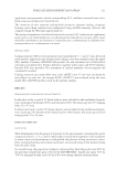
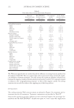
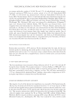
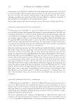
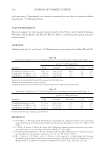
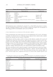
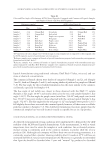
JOURNAL OF COSMETIC SCIENCE 230 tical exposures. Consequently, we cannot recommend porcine skin for routine mildness screening by 14 C-SDS penetration. ACKNOWLEDGMENTS Financial support for this research was provided by the Procter and Gamble Company. We thank Tiffany Brooks and Keun Il Kim for help in conducting the human skin pen- etration assays. APPENDIX Tabular results for 10- and 2-min 14 C-SDS penetration into human skin (Tables III and IV). REFERENCES (1) P. N. Moore, S. Puvvada, and D. Blankschtein, Challenging the surfactant monomer skin penetration model: Penetration of sodium dodecyl sulfate micelles into the epidermis, J. Cosmet. Sci., 54(1), 29–46 (2003). (2) C. Froebe, F. Simion, L. Rhein, R. Cagan, and A. Kligman, Stratum corneum lipid removal by surfac- tants: Relation to in vivo irritation, Dermatology, 181(4), 277–283 (1990). (3) S. Ghosh and D. Blankschtein, The role of sodium dodecyl sulfate (SDS) micelles in inducing skin bar- rier perturbation in the presence of glycerol, J. Cosmet. Sci., 58, 109–133 (2007). (4) M. J. Fevola, R. M. Walters, and J. J. LBrizzi, A new approach to formulating mild cleansers: Hydrophobically-modified polymers for irritation mitigation, in Polymeric Delivery of Therapeutics, S. E. Morgan and R. Y. Lochhead, eds. (American Chemical Society, New York, 2010), pp. 221–242. Table IV Treatments, Sample Sizes, and 14 C-SDS Disposition for 2-min Exposure on Human Skin (Expt. 2) Treatment μg/cm2 14 C-SDS equivalents Wipe Receptor Penetration (+) SE (-) SE 50 mM SLS (control) (n = 20) 3.2 ± 0.6 0.06 ± 0.04 7.3 1.2 1.0 50 mM SLS + 2% PEO (n = 21) 3.1 ± 1.5 0.07 ± 0.05 3.5 0.5 0.5 50 mM SLS + 2% PVA (n = 21) 3.2 ± 0.4 0.11 ± 0.07 5.3 0.8 0.7 Columns have the same meaning as in Table III. Table III Treatments, Sample Sizes, and 14 C-SDS Disposition for 10-min Exposure on Human Skin (Expt. 1) Treatment μg/cm2 14 C-SDS equivalents Wipea Receptorb Penetrationc (+) SE (-) SE 50 mM SLS (control) (n = 14) 8.0 ± 1.9 0.03 ± 0.01 16.0 2.3 2.0 50 mM SLS + 2% PEO (n = 15) 2.8 ± 0.6 0.05 ± 0.03 6.5 0.8 0.7 50 mM SLS + 2% PVA (n = 14) 12.9 ± 7.0 0.09 ± 0.05 11.3 1.5 1.3 a Radioactivity removed with wetted fi lter papers after the wash step. b Radioactivity measured in receptor solution. c Geometric mean of the total radioactivity found in all layers of skin + receptor solution.
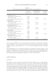
Purchased for the exclusive use of nofirst nolast (unknown)
From: SCC Media Library & Resource Center (library.scconline.org)