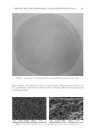
JOURNAL OF COSMETIC SCIENCE 260 room condition was controlled at 23 ± 2°C with 40–70% relative humidity. The ultimate tensile strength and percent elongation at break value were calculated by these formulas: Ultimatetensilestrength(kg/mm2) breakingload cross-sectionareaof thetestedspecimen Percentelongationatbreak Differentinthelengthatbreakingpoint 100 originallengthof thetestedspecimen = = × MOISTURE CONTENT Moisture analyzer (Sartorius Model MA30, DKSH GmbH, Hamburg, Germany) was used to measure the percentage of moisture content of the formulated patches. The tested patches were cut in 0.2 × 0.2 cm2 and approximately 5 g was placed in a preweighted aluminum dish. The dishes and contents were put in an oven at 105°C for 12 min and then placed in a desiccator to cool down before weighing. The percentage of moisture content was calculated from the below formula: % Moisturecontent Initialweight Finalweight 100 Initial weight = - × The experiment was performed in triplicate. Surface morphology. The surface morphology of the formulated patch was determined by using a scanning electron microscope (SEM Model 1455VP LEO, Cambridge, UK). The tested patches were dried in a hot-air oven at 40°C and kept in a desiccator to constant weight before testing. Double-sided tape was used to keep the dried patch attached on the stub. All tested patches on stub were coated with gold. All images were manifested at 350 times. IN VITRO RELEASE STUDY OF ARTOCARPIN FROM THE FORMULATED PATCH INCORPORATING THE EXTRACT In vitro release of artocarpin from the formulated patches was evaluated by using the vertical Franz diffusion cell (Model V6A-02/90824 PermeGear Inc., Hellertown, PA). The area of the tested patch exposed to the phosphate buffer (pH 7.4), the receptor medium, was 2.27 cm2. The volume of the phosphate buffer in the receptor chamber was 12 mL with the temperature set at 37°C. This whole assembly was kept on a magnetic stirrer, and the receptor medium was stirred continuously using a magnetic bead. The receptor medium were collected at various time intervals (5, 10, 15, 30, 60,120, and 240 min) and replaced with an equal volume of fresh medium. The amount of artocarpin that diffused through the patch and accumulated in the receptor medium was determined by HPLC. The study was run in triplicate. EFFICACY AND TOLERANCE STUDY OF THE FORMULATED PATCH INCORPORATING THE EXTRACT The study protocol was approved by Human Ethical Committee, Naresuan University, Phitsanulok, Thailand with the permission number 54 03 03 0004. All procedures were

CHITOSAN PATCH INCORPORATING A. ALTILIS HEARTWOOD EXTRACT 261 performed at Cosmetics and Natural Products Research Center, Naresuan University, Phitsanulok, Thailand. Selection criteria for subjects. Healthy Thai male or female with age 20–45 year and having a melanin value, in a hyperpigmented area on facial skin, in a range of 290–490 AU were included. Subjects were excluded from the study if they had been smoking and alcoholic. Female subjects were excluded if they were pregnant or lactating. Subjects were to have discontinued topical application of any medicines, steroids, photosensitizing, or cosmetics with depigmenting agents such as tretinoin, α-hydroxy acids, β-hydroxy acids, hydro- quinone, kojic acid, and arbutin on facial skin for at least 2 weeks before study. In addi- tion, subjects were not to have used any systemic steroids, hormones, antibiotics, and antihistamines for at least 4 weeks before the study. Other exclusion criteria included subjects who had a scar or burn with a diameter of more than 2 cm per each area on face or forearm, or who had history of atopic skin reaction, eczema, psoriasis, or recurrent or active herpes simplex on face or forearm. Excessive exposure to sunlight was to be avoided. Irritation test. Subjects were screened according to inclusion and exclusion criteria. Only subjects who met the criteria entered the skin irritation test (4-h patch test), which was designed to assess the skin tolerance to the formulated patch. All subjects were asked to sign an informed consent before screening into the study. To assess the irritant contact dermatitis, the chitosan hydrogel patch incorporating the extract (the tested patch, 4 cm2 of patch containing 0.29 mg of artocarpin), 20% sodium lauryl sulfate (SLS BASF (Thai) Ltd., Bangkok, Thailand) (positive control, 0.2 ml), and distilled water (negative control, 0.2 ml) were tested using the 4-h human patch test. The testing samples were applied to the three areas on subject’s forearm. Each area was sepa- rated by 3 cm2 from each other, then covered with a Webril pad for up to 4 h. The test patch and substances were removed or wiped off from the skin. Treatment sites were as- sessed for the presence of irritation at 0.5, 24, 48, and 72 h after patch test removal. The evaluation of skin irritation was composed of objective assessment using Mexameter® (Model MX 18 Courage and Khazaka Electronic GmbH, Cologne, Germany) for skin redness measurement (erythema index) and visual assessment by a dermatologist in three domains including erythema, scaling, and oedema, according to the scale of Frosch & Kligman and COLIPA (9,12,13). Effi cacy test. The study design was a randomized, double-blind and parallel study. The site of testing was the facial skin. The subjects were randomly assigned to either receive the test patch (size of 4 cm2 of the formulated patch containing 0.29 mg of artocarpin, test group) or the control patch (size of 4 cm2 of the formulated patch without the extract, control group) applying it to the hyperpigmented area. The control patch had components simi- lar to the test patch but without the extract. To determine the balance in skin properties of the two groups, their skin properties at the hyperpigmented area were evaluated before (baseline, week 0) and after patch application. Duration of the study was 8 weeks. At week 0 of the study, subjects arrived at the testing room at 8.00 am. They were asked to wash their non–make up face with clean water, pat the face dry with a towel, and wait for 30 min before proceeding to the next procedure of measuring the skin properties. The temperature and humidity of the test room were controlled to 25 ± 2°C and 50–70% RH, respectively. Skin properties including skin colors (melanin and erythema values), moisture content and skin pH of the hyperpigmented area were measured by using Mexameter®, Corneometer® (Model CM285 Courage and Khazaka Electronic GmbH) and
Purchased for the exclusive use of nofirst nolast (unknown)
From: SCC Media Library & Resource Center (library.scconline.org)