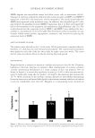
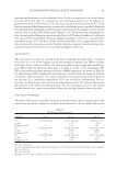
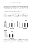
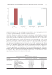
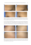
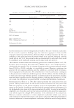
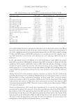
JOURNAL OF COSMETIC SCIENCE 62 product was used until 8 weeks after the treatment (Table III). Conversely, the test cream produced a statistically signifi cant decrease in eye wrinkles after 4 weeks of use. In addi- tion, the results show that there were signifi cant differences between the test and placebo creams in Rz and Rt parameters after 8 weeks of treatment. Our fi ndings indicate that the test cream smoothed the skin surface and reduced wrinkles. CONCLUSIONS This study was carried out to evaluate the antiaging effect of PT-FX50. The results show that PT-FX50 is not cytotoxic at concentrations of d20 ppm. We have also shown that PT-FX50 decreases the expression levels of MMP-1, MMP-2, and MMP-9 in HDFN cells. Furthermore, the fucoxanthin-containing cream we prepared showed considerable effi cacy as it signifi cantly increased skin moisture and elasticity after 4 weeks of treat- ment. These results demonstrate that the test product has excellent antiwrinkle and moisturizing effects. Furthermore, our fi ndings suggest that fucoxanthin can be used as a functional cosmetic agent. A CKNOWLEDGMENT T his research was a part of the project titled “Development and industrialization of high value cosmetic raw materials from marine microalgae,” funded by the Ministry of Oceans and Fisheries, Korea. R EFERENCES ( 1) M. A. Farage, K. W. Miller, P. Elsner, and H. I. Maibach, Intrinsic and extrinsic factors in skin ageing: a review, Int. J. Cosmet. Sci., 30, 87–95 (2008). ( 2) H. K. Biesalski, M. Berneburg, T. Grune, M. Kerscher, J. Krutmann, W. Raab, J. Reimann, T. Reuther, L. Robert, and T. Schwarz, Hohenheimer Consensus Talk. Oxidative and premature skin ageing, Exp. Dermatol., 12(Suppl. 3), 3–15 (2003). Table III Evaluation of Skin Wrinkles Parameters Day 0 Day 28 Day 56 Rab Test 5.295 ± 0.920 5.062 ± 0.980 5.016 ± 0.960 Placebo 5.258 ± 1.053 5.209 ± 1.005 5.105 ± 0.834 p valuea — 0.140 0.413 Rzc Test 32.871 ± 6.221 30.507 ± 4.655 28.633 ± 4.701 Placebo 31.499 ± 5.479 30.690 ± 5.883 30.056 ± 4.093 p value — 0.149 0.034 Rtd Test 46.110 ± 8.781 41.578 ± 6.516 38.630 ± 5.569 Placebo 44.183 ± 7.821 42.935 ± 9.084 41.356 ± 4.807 p value — 0.123 0.037 a p value: signifi cant probability, linear mixed model, p 0.05 when test and placebo products are compared. b Ra: arithmetic average roughness. c Rz: arithmetic average of the difference segment roughness calculated from fi ve succeeding measurements. d Rt: the distance between the highest mountain and the lowest value.

ANTIAGING POTENTIAL OF FUCOXANTHIN 63 ( 3) H. W. Daniell, Smoker’s wrinkles. A study in the epidemiology of “crow’s feet”, Ann. Intern. Med., 75, 873–880 (1971). ( 4) G. L. Grove, M. J. Grove, and J. J. Leyden, Optical profi lometry: an objective method for quantifi cation of facial wrinkles, J. Am. Acad. Dermatol., 21, 631–637 (1989). ( 5) C. E. Griffi ths, T. S. Wang, T. A. Hamilton, J. J. Voorhees, and C. N. Ellis, A photonumeric scale for the assessment of cutaneous photodamage, Arch. Dermatol., 128, 347–351 (1992). ( 6) C. Y. Lu, H. C. Lee, H. J. Fahn, and Y. H. Wei, Oxidative damage elicited by imbalance of free radical scavenging enzymes is associated with large-scale mtDNA deletions in aging human skin, Mutat. Res., 423, 11–21 (1999). ( 7) S. J. Moloney, S. H. Edmonds, L. D. Giddens, and D. B. Learn, The hairless mouse model of photoaging: evaluation of the relationship between dermal elastin, collagen, skin thickness and wrinkles, Photochem. Photobiol., 56, 505–511 (1992). ( 8) S. K. Moon, S. K. Kang, and C. H. Kim, Reactive oxygen species mediates disialoganglioside GD3- induced inhibition of ERK1/2 and matrix metalloproteinase-9 expression in vascular smooth muscle cells, FASEB J., 20, 1387–1395 (2006). ( 9) K. K. Nelson and J. A. Melendez, Mitochondrial redox control of matrix metalloproteinases, Free Radic. Biol. Med., 37, 768–784 (2004). ( 10) L. Grange, M. V. Nguyen, B. Lardy, M. Derouazi, Y. Campion, C. Trocme, M. H. Paclet, P. Gaudin, and F. Morel, NAD(P)H oxidase activity of Nox4 in chondrocytes is both inducible and involved in collage- nase expression, Antioxid. Redox. Signal., 8, 1485–1496 (2006). ( 11) M. H. Shin, Y. J. Moon, J. E. Seo, Y. Lee, K. H. Kim, and J. H. Chung, Reactive oxygen species pro- duced by NADPH oxidase, xanthine oxidase, and mitochondrial electron transport system mediate heat shock-induced MMP-1 and MMP-9 expression, Free Radic. Biol. Med., 44, 635–645 (2008). ( 12) J. S. Weiss, C. N. Ellis, J. T. Headington, T. Tincoff, T. A. Hamilton, and J. J. Voorhees, Topical treti- noin improves photoaged skin. A double-blind vehicle-controlled study, JAMA, 259, 527–532 (1988). ( 13) R. Hermitte, Aged skin, retinoids and alpha hydroxy acids, Cosmet. Toilet., 107, 63–67 (1992). ( 14) D. S. Rosenthal, D. R. Roop, C. A. Huff, J. S. Weiss, C. N. Ellis, T. Hamilton, J. J. Voorhees, and S. H. Yuspa, Changes in photo-aged human skin following topical application of all-trans retinoic acid, J. Invest. Dermatol., 95, 510–515 (1990). ( 15) C. M. Ditre, T. D. Griffi n, G. F. Murphy, H. Sueki, B. Telegan, W. C. Johnson, R. J. Yu, and E. J. Van Scott, Effects of alpha-hydroxy acids on photoaged skin: a pilot clinical, histologic, and ultrastructural study, J. Am. Acad. Dermatol., 34, 187–195 (1996). ( 16) A. El-Agamey, G. M. Lowe, D. J. McGarvey, A. Mortensen, D. M. Phillip, T. G. Truscott, and A. J. Young, Carotenoid radical chemistry and antioxidant/pro-oxidant properties, Arch. Biochem. Biophys., 430, 37–48 (2004). ( 17) J. Peng, J. P. Yuan, C. F. Wu, and J. H. Wang, Fucoxanthin, a marine carotenoid present in brown seaweeds and diatoms: metabolism and bioactivities relevant to human health, Mar. Drugs, 9, 1806– 1828 (2011). ( 18) R. Pangestuti and S. K. Kim, Biological activities and health benefi t effects of natural pigments derived from marine algae, J. Funct. Foods, 3, 255–266 (2011). ( 19) C. S. Kumar, P. Ganesan, P. V. Suresh, and N. Bhaskar, Seaweeds as a source of nutritionally benefi cial compounds - a review, J. Food Sci. Technol., 45, 1–13 (2008). ( 20) M. Hosokawa, T. Okada, N. Mikami, I. Konishi, and K. Miyashita, Bio-functions of marine carot- enoids, Food Sci. Biotechnol., 18, 1–11 (2009). ( 21) S. J. Heo, W. J. Yoon, K. N. Kim, G. N. Ahn, S. M. Kang, D. H. Kang, A. Affan, C. Oh, W. K. Jung, and Y. J. Jeon, Evaluation of anti-infl ammatory effect of fucoxanthin isolated from brown algae in lipo- polysaccharide-stimulated RAW 264.7 macrophages, Food Chem. Toxicol., 48, 2045–2051 (2010). ( 22) A. Jiménez-Escrig, I. Jiménez-Jiménez, R. Pulido, and F. Saura-Calixto, Antioxidant activity of fresh and processed edible seaweeds, J. Sci. Food Agric., 81, 530–534 (2001). (23 ) I. Urikura, T. Sugawara, and T. Hirata, Protective effect of fucoxanthin against UVB-induced skin pho- toaging in hairless mice, Biosci. Biotechnol. Biochem., 75, 757–760 (2011). (24 ) N. M. Sachindra, E. Sato, H. Maeda, M. Hosokawa, Y. Niwano, M. Kohno, and K. Miyashita, Radical scavenging and singlet oxygen quenching activity of marine carotenoid fucoxanthin and its metabolites, J. Agric. Food Chem., 55, 8516–8522 (2007). (25 ) F. Beppu, M. Hosokawa, M. J. Yim, T. Shinoda, and K. Miyashita, Down-regulation of hepatic stearoyl- CoA desaturase-1 expression by fucoxanthin via leptin signaling in diabetic/obese KK-A(y) mice, Lip- ids, 48, 449–455 (2013).
Purchased for the exclusive use of nofirst nolast (unknown)
From: SCC Media Library & Resource Center (library.scconline.org)