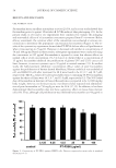
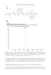
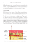
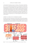
JOURNAL OF COSMETIC SCIENCE 102 which induces the electrostatic interaction between the zein protein and SDS. In addi- tion, the sizes of polar heads of nonionic surfactants or certain amphoteric surfactants could increase the steric hindrance, thereby reducing the access of the hydrophobic side chains of proteins into internal micelles and the exposure of hydrophobic parts of surfac- tant micelles to the external environment (48). THE PENETRATION OF SURFACTANTS INTO SKIN As discussed earlier, one reason to cause the surfactant-induced skin irritation is the con- tact between surfactants and the lipids/proteins in the skin barrier. If the surfactant pen- etration into the skin can be inhibited, its contact with skin lipids/proteins can be reduced, so is the surfactant-induced skin irritation. To achieve this, the understanding in the mechanism of the surfactant penetration into the skin is essential. In the past two decades, many scientists have researched this topic. To summarize, there has been mainly three hypotheses respecting the mechanism of surfactant penetration into the skin. THE SURFACTANT MONOMER PENETRATION MODEL A widely accepted view of the surfactant penetration is termed as the monomer penetration model. This model explains that surfactant monomers can access the pathways through the skin barrier because they possess relatively small sizes. When the surfactant monomers penetrate into the skin barrier, they interact with skin proteins and lipids, inducing skin irritation. On the other hand, when the concentration of surfactant monomers reaches the CMC, micelles are formed, which have relatively larger sizes and lower surface activity (50). Consequently, they lack the ability to penetrate into the skin barrier. The monomer penetration model had been investigated by many researchers, including Ghosh and Blankschtein (51). Sodium cocoyl isethionate (SCI) was studied because it was considered as a mild surfactant. Past studies demonstrated that SCI did not induce serious irritation compared with other anionic surfactants (52). Ghosh and Blankschtein (51) recorded the shift of skin electrical current versus SCI concentration, which did not change further beyond the CMC of SCI. This clearly indicated that the SCI micelles lack the ability to penetrate into the skin. THE SURFACTANT MICELLE AND THE SUBMICELLE PENETRATION MODEL The surfactant monomer penetration model suggests that the penetration of the surfac- tant into the skin is dose independent—the amount of surfactants presented in the skin would not increase further when the surfactant concentration exceeds the CMC. However, skin penetration of SDS showed contradicting results. Moore et al. (50) studied the rela- tionship between the SDS concentration and the amount of SDS penetrating into the skin. The results demonstrated that when the concentration of SDS was beyond the CMC, the amount of SDS in the skin barrier still increased without any limitation. As a result, the penetration behavior of SDS could not be simply explained by the monomer penetra- tion model (53). Through the experiments directed by Ghosh and Blankschtein (51), they found out that the SDS micelles had the ability to penetrate into the skin. Moreover, SDS skin penetration beyond the CMC was predominated by the SDS micelles. This is proposed as the surfactant micelle penetration model.

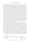
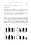
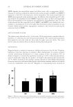
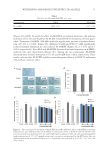
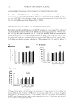
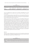
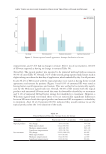
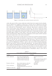
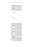
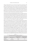
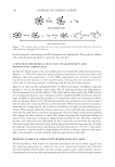
SURFACTANT PENETRATION 103 Clearly, contradicted behaviors were observed from the penetration experiments of SCI and SDS. Consequently, researchers did a series of studies and proposed hypotheses to perfect the surfactant penetration model. The CMC of SCI is indeed lower than that of SDS, inducing a smaller amount of monomers existing in the solution and the formation of micelles at a lower concentration (51). According to the monomer penetration model, a surfactant with a lower CMC is unlikely to cause high skin penetration and induce seri- ous irritation (54), which partially explains why SCI is milder than SDS (55). But how about the penetration behaviors of SCI micelles? To answer this question, Ghosh and Blankschtein (51) measured the radius of SCI micelles and the average aqueous pore ra- dius resulting from skin exposure to SCI by using dynamic light-scattering. The results as well as their comparison to SDS and control are presented in Table IV. Apparently, SCI micelles are larger than SDS micelles, which were also larger than the aver- age aqueous pore radius. This indicated that SCI micelles face steric hindrance to penetrate into the skin barrier. On the other hand, SDS micelles are small enough to be able to enter the skin aqueous pores. Therefore, micelle size is an important factor to determine the sur- factant penetration behaviors. Moreover, SDS is capable of increasing both the aqueous pore size and the number density thus, it could alter the skin structure, inducing skin irritation (51). Ghosh and Blankschtein (51) demonstrated that SCI only slightly induced skin per- turbation, thanks to its large micelle size and its ability to reduce aqueous pore radius/ density. As a result, SCI is a good candidate to be applied to mild cleansing. Hill et al. (56) and James-Smith et al. (57,58) proposed the submicelle penetration model. This hypothesis was based on micelle kinetics—micelles are rapidly breaking and reform- ing continuously (57–59). This dynamic state is described in Figure 7. The dynamic state includes two types of relevant time scales: fast relaxation time and slow relaxation time. Fast relaxation time is used to measure the time needed for the surfactants to enter or come out of the micelles. Slow relaxation time describes the time used for the micelles to completely form or completely integrate. In this equilibrium, some monomers could form aggregations smaller than micelles termed submicelles or premicelles. James-Smith et al. (58) observed SDS submicelles were presented at the concentration of 3–4 times CMC. Later, LeBard et al. (59) used the molecular dynamic simulations to investigate the dynamic change of a nonionic polyethylene glycol (PEG) surfactant, C7E6 solution, at low concentrations. Pre- micelles were identifi ed at concentrations below the CMC, and their concentration increased with increasing total concentration below the CMC, reaching a plateau above the CMC, where these premicelles exist in equilibrium with free monomers and full-size micelles (59). Because of the smaller sizes, the submicelles may have the ability to penetrate into the skin barrier (56–58). To verify this hypothesis, Hill et al. (57) adjusted the SDS micelle stability by mixing with dodecyl trimethylammonium bromide (C12TAB) at various ratios and ob- served that addition of C12TAB lowered the ability of SDS to perturb skin barrier properties Table IV Micelle Radius and the Average Aqueous Pore Radius after Skin Exposure to SCI/SDS/Phosphate-buffered saline (PBS) Control Solutions Types of solution Micelle radius, r (A) Average aqueous pore radius, rpore (A) Aqueous pore number density, (ε/τ)normal SCI solution 33.5 ± 1 29 ± 5 2 ± 1 SDS solution 19.5 ± 1 33 ± 5 7 ± 1 PBS control solution Not applicable 20 ± 3 1
Purchased for the exclusive use of nofirst nolast (unknown)
From: SCC Media Library & Resource Center (library.scconline.org)