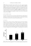
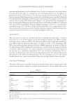
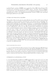
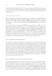
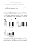
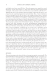
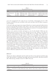
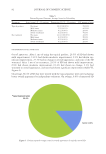
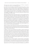
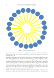
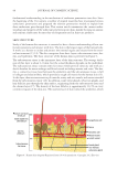
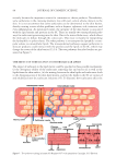
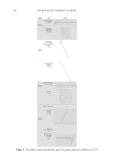
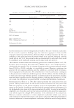
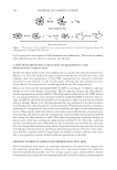
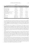
JOURNAL OF COSMETIC SCIENCE 104 by decreasing the concentration of SDS monomers and submicelles. This indicates submi- celles indeed possess the ability to penetrate into the skin. A NEW PROPOSED MODEL: SURFACTANT CHARGE DENSITY AND PENETRATION CORRELATION To fi nd out which model is the most sophisticated to explain the surfactant penetration, Morris et al. (60) well studied the physicochemical parameters of surfactants that could infl uence their skin penetration: (i) the CMC, representing the amount of surfactant monomers in the solution (ii) the micelle radius, showing the steric hindrance of mi- celles and (iii) the zeta potential, correlating with the micelle charge density. Morris et al. (60) used the radiolabeled SDS (14C-SDS) to investigate 16 different surfactant systems in vitro on the human cadaver skin. The 16 surfactant systems and their physico- chemical parameters are listed in Table V. The study indicated that only (i) the CMC and (iii) the zeta potential showed a clear correlation with the radiolabeled SDS penetration. Interest- ingly, the SDS micelle radius did not show a clear correlation, which was inconsistent with the previous fi ndings. Morris et al. (60) hypothesized this was attributed to the short residence time of surfactants contacting the skin in the experiment. When the skin barrier is exposed to surfactants for a longer period, the micelle size would have an impact on the surfactant pene- tration. When anionic surfactant solution comes into contact with the skin, monomer pene- trates the skin and binds to proteins, increasing the electrical charge on the protein network and causing the skin structure to swell. This allows for progressive surfactant binding in deeper layers of the skin, resulting in enhanced skin swelling (57,61). Therefore, surfactant micelles and submicelles may additionally be able to penetrate and swell the skin structure. The more charged the surfactant system and the longer the surfactant exposure time, the more the binding to the skin proteins, speeding up the penetration process (33,34,57). Zeta poten- tial is known to correlate with the charge density of colloids in solution, and that is the reason why it was revealed to correlate with the surfactant penetration in the study. METHODS TO REDUCE SURFACTANT PENETRATION INTO SKIN All the hypotheses with respect to surfactant penetration discussed earlier suggest that the surfactant penetration is related to the steric interaction between the surfactant monomers/submicelles/micelles and the average aqueous pore radius/number density. Consequently, increasing surfactant monomer/micelle size and/or reducing average aqueous Figure 7. The dynamic state of surfactants at a certain concentration of surfactants (Reprinted from (58) with permission. Copyright 2007 Elsevier).

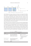
SURFACTANT PENETRATION 105 pore radius/number density could play an important role in skin barrier protection. Bind- ing surfactants with other bulky molecules such as nonionic surfactants and polymers is one of the common methods to increase surfactant micelle size (62). For example, mixing C12E6 with SDS reduces SDS CMC and induces its micelle growth (63), thereby limit- ing SDS monomer and micelle skin penetration and reducing SDS-induced skin irritation. In the experiments directed by Moore et al. (50), Polyethylene oxide (PEO) was mixed with SDS in solution. Hydrophilic polymers like PEO are known to form micelle-like complexes with anionic surfactants such as SDS, with the polymer forming a corona around the anionic surfactant micelles. An obvious increase in the average SDS micelle radius from 20 to 25 A was observed with the addition of PEO. This binding effectively reduced SDS skin penetration attributed to the steric hindrance and/or the slow diffusion of SDS. Adding humectant to the surfactant solution is known to improve the skin mildness by providing hydration (64,65). Ghosh and Blankschtein (66) demonstrated that it is also an effective way to reduce surfactant penetration. In their study (66), electrical currents served as an index to refl ect the amount of SDS presented in the skin barrier. The results indicated that the amount of SDS in the skin continuously increased when the pure SDS concentration exceeded the CMC. However, it was not the case for the mixture of SDS in 10% glycerol solution. Ghosh and Blankschtein (66) stated three hypothesizes to account for the reduced SDS penetration by adding 10% glycerol: (i) the glycerol addition reduced the SDS CMC (ii) the addition of the 10% glycerol increased the SDS micelle size, hindering them from penetrating into the skin (iii) 10% glycerol reduced the radius and/or the density of the aqueous pores in the skin barrier. Both (i) and (ii) hypotheses were proved to be invalid. The data in Table VI demonstrated that the 10% glycerol addition effectively reduced the average pore radius and the pore number density in the skin. Table V CMCs, Micelle Diameter, and Zeta Potential of 16 Surfactant Systems Tested by 14 C-SDS Surfactant system CMC, mM Micelle diameter, nm Zeta potential, mV SLS 3.15 ± 0.03 2.73 ± 0.19 -54.1 ± 3.8 SLS in NaCl (0.01 M) 2.64 ± 0.31 3.43 ± 0.16 — SLS in NaCl (0.05 M) 1.43 ± 0.12 4.86 ± 0.26 — SLS in NaCl (0.10 M) 1.06 ± 0.07 5.51 ± 0.15 — SLS in NaCl (0.25 M) 0.671 ± 0.0015 0.628 ± 0.06 — SLS with 2% PEO 2.30 ± 0.18 3.04 ± 0.12 -24.2 ± 0.7 SLS with Dimethyl dodecyl amine oxide 4.86 ± 0.22 126 ± 20 -77.5 ± 3.6 SLS with Lauramidopropyl betaine 0.526 ± 0.032 4.10 ± 0.30 -52.9 ± 6.5 SLS with Lauric acid 9.26 ± 0.08 2.26 ± 0.10 -72.7 ± 4.1 Sodium dodecyl benzene sulphonate 2.00 ± 0.08 3,94 ± 0.07 -47.9 ± 5.1 SLE1 S 1.32 ± 0.04 2.26 ± 0.15 -46.1 ± 4.5 SLE3S 0.452 ± 0.037 2.19 ± 0.06 -27.6 ± 2.3 SLI with LAPB 0.289 ± 0.023 4.07 ± 0.41 -63.0 ± 6.3 C12E6 0.0695 ± 0.0014 25.3 ± 1.1 -7.2 ± 0.9 SLS with C12E5 0.0993 ± 0.0022 1.85 ± 0.06 -39.4 ± 4.7 SLS with C12E6 0.0992 ± 0.0034 1.91 ± 0.17 -32.5 ± 1.4
Purchased for the exclusive use of nofirst nolast (unknown)
From: SCC Media Library & Resource Center (library.scconline.org)