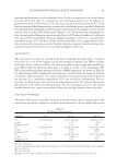
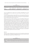
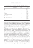
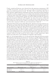
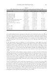
JOURNAL OF COSMETIC SCIENCE 106 CONCLUSION AND PERSPECTIVES The focus of this article is to summarize the state-of-the-art understanding of surfac- tants’ penetration into the skin, which have been studied by many researchers for decades. Nevertheless, an explicit surfactant penetration model still could not be given so far. It is likely that different penetration hypotheses play a role simultaneously, and a combi- nation of all the mechanisms enables surfactant penetration into the skin. With respect to mild cleansing, the addition of nonionic/amphoteric surfactants, hydrophilic polymers, or humectants such as glycerol can minimize the skin penetration of anionic surfactants, reducing the occurrence of skin irritation. On the other hand, a complete prevention of surfactant penetration into the skin is diffi cult and challenging, implying that surfactants can be used as penetration enhancers for transepidermal-active delivery. ACKNOWLEDGMENTS The author is grateful for the opportunities provided by the College of Pharmacy, Shenyang Pharmaceutical University, and the ViaX Online Education. REFERENCES (1 ) K. Holmberg, B. Jönsson, B. Kronberg, and B. Lindaman, “Introduction of surfactants,” in Surfactants and Polymers in Aqueous Solution, 2nd Ed., John Wiley & Sons Ltd., (2002), pp. 1–23. (2) A. Mehling, M. Kleber, and H. Hensen, Comparative studies on the ocular and dermal irritation poten- tial of surfactants. Food Chem. Toxicol., 45, 747–758 (2007). (3) Y. Nakama, “Sufactants,” in Cosmetic Science and Technology, 1st Ed., Elsevier Ltd., (2017), pp. 231–224. (4 ) A. Seweryn, Interactions between surfactants and the skin – theory and practice, Adv. Colloid Interf. Sci., 256, 242–255 (2018). ( 5) L. Zhang, X. Zhang, P. Zhang, Z. Zhang, S. Liu, and B. Han, Effi cient emulsifying properties of glyc- erol-based surfactant, Colloids Surf. A, 553, 225–229 (2018). (6 ) P. López-Mahía, S. Muniategui, D. Prada-Rodríguez, and M. C. Prieto-Blanco, “Surfactants and deter- gents,” in Encyclopedia of Analytical Science, 2nd Ed. Elsevier Ltd., (2005), pp. 554–561. (7) D. Bajpai, A. Mishra, J. Clark, T. Farmer, Synthesis, chemistry, physicochemical properties and indus- trial applications of amino acid surfactants: a review, Compt. Rendus Chem., 21, 112–130 (2018). (8) Y . Yu, J. Zhao, and A. E. Bayly, Development of surfactants and builders in detergent formulations. Chin. J. Chem. Eng., 16(4), 517–527 (2008). (9) J . Steber, “The ecotoxicity of cleaning product ingredients.” in Handbook for Cleaning/Decontamination of Surfaces., 1st Ed., Elsevier B.V, (2007), Vol. 2, pp. 721–746. (10) D . Zhao and Y. Wan, The synthesis of mesoporous molecular sieves. Stud. Surf. Sci. Catal., 168, 241– 300 (2007). (11) M . Teresa Garcia, E. Campos, A. Marsal, and I. Ribosa, Fate and effects of amphoteric surfactants in the aquatic environment, Environ. Int., 34, 1001–1005 (2008). Table VI Skin Aqueous Pore Radius and Normalized Pore Number Density Resulted by Various Solutions Solution Average pore radius, rpore (A) Normalized pore number density, (ε/τ)normal SDS 33 ± 5 7 ± 1 SDS with 10% glycerol 20 ± 5 3 ± 1 PBS control 20 ± 3 1 10% glycerol 11 ± 4 0.5 ± 0.1 See references 68., 69., 70., 71., 72., 73.

SURFACTANT PENETRATION 107 (12) J . Escalas-Taberner, E. González-Guerra, and A. Guerra-Tapia, Sensitive skin: a complex syndrome, Actas Dermosifi liogr, 102(8), 563–571 (2011). (13) G . Honari and H. Maibach, “Skin structure and function,” in Applied Dermatotoxicology., 1st Ed., Elsevier Inc., (2014), pp. 1–10. (14) J. G. Marks, Jr. and J. J. Miller, “Structure and function of the skin,” in Lookingbill and Marks’ Principles of Dermatology, 6th Ed. Elsevier Inc., (2019), pp. 2–10. (15) S. Nafi si and H. I. Maibach, “Skin penetration of nanoparticles,” in Emerging Nanotechnologies in Immunol- ogy., 1st Ed., Elsevier Inc., (2018), pp. 47–88. (16) K. Van der Maaden, W. Jiskoot, and J. Bouwstra, Microneedle technologies for (trans) dermal drug and vaccine delivery, J. Contr. Release, 161, 645–655 (2012). (17) A. M. Hargis and S. Myers, “The integument,” in Pathologic Basis of Veterinary Disease, 6th Ed. Elsevier Inc., (2017), pp. 1009–1146. (18) H . SchlÜter, E. Upjohn, G. Varigos, and P. Kaur, Burns and skin ulcers,” in Essentials of Stem Cell Biol- ogy, 3rd Ed. Elsevier Inc., (2014), 501–513. (19) J. - H. Lee, “Keratinocyte differentiation and epigenetics,” in Epigenetics and Dermatology., 1st Ed., Elsevier Inc., (2015), pp. 37–52. (20) S. Chermprapai, F. Broere, G. Gooris, Y. M. Schlotter, V. P. M. G. Rutten, and J. A. Bouwstra, Altered lipid properties of the stratum corneum in canine atopic dermatitis, Biochim. Biophys. Acta Biomembr., 1860, 526–533 (2018). (21) N. Dayan, “Delivery system design in topically applied formulations: an overview,” in Delivery System Handbook for Personal Care and Cosmetic Products. (2015), pp. 101–118. (22) K. P. Ananthapadmanabhan, S. Mukherjee, and P. Chandar, Stratum corneum fatty acids: their critical role in preserving barrier integrity during cleansing, Int. J. Cosmet. Sci., 35, 337–345 (2013). (23) P. W. Wertz and B. van den Bergh, The physical, chemical, and functional properties of lipids in the skin and other biological barriers, Chem. Phys. Lipids, 91, 85–96 (1998). (24) N. Nakagawa, S. Sakai, M. Matsumoto, K. Yamada, M. Nagano, T. Yuki, Y. Sumida, and H. Uchiwa, Relationship between NMF (lactate and potassium) content and the physical properties of the stratum corneum in healthy subjects, Soc. Invest. Dermatol., 122, 755–763 (2004). (25) S. BiÖrklund, J. Engblom, K. Thuresson, and E. Sparr, Glycerol and urea can be used to increase skin permeability in reduced hydration conditions, Eur. J. Pharm. Sci., 50, 638–645 (2013). ( 26) A. Watabe, T. Sugawara, K. Kikuchi, K. Yamasaki, S. Sakai, and S. Aiba, Sweat constitutes several natural moisturizing factors, lactate, urea, sodium, and potassium, J. Dermatol. Sci., 72, 177–182 (2013). ( 27) J. A. Bouwstra, G. S. Gooris, J. A. van der Spek, W. Bras, Structural investigations of human stratum corneum by small-angle X-ray scattering, Soc. Invest. Dermatol., 97, 1005–1012 (1991). ( 28) B. Forslind, A domain mosaic model of the skin barrier, Acta Derm. Venereol., 74, 16 (1994). ( 29) R. M. Wang, S. R. Zheng, and Y. P. Zheng, “Reinforced materials,” in Polymer Matrix Composites and Technology. (2011), pp. 527–528. ( 30) K. R. Feingold, M. Schmuth, and P. M. Elias, The regulation of permeability barrier homeostasis, J. Invest. Dermatol., 127, 1574–1576 (2007). ( 31) Y. Kitajima, Implications of normal and disordered remodeling dynamics of corneodesmosomes in stra- tum corneum, Dermatol. Sin., 33, 58–63 (2015) ( 32) M. Gelker, C. C. Müller-Goymann, W. ViÖl, Permeabilization of human stratum corneum and full- thickness skin samples by a direct dielectric barrier discharge, Clin. Plasma Med., 9, 34–40 (2018). (3 3) K. P. Ananthapadmanabhan, D. J. Moore, K. Subramanyan, M. Misra, and F. Meyer, Cleansing without compromise: the impact of cleansers on the skin barrier and the technology of mild cleansing, Dermatol. Ther., 17, 16–25 (2004). (3 4 ) E. Lémery, S. Briancon, Y. Chevalier, C. Bordes, T. Oddos, A. Gohier, and M.-A. Bolzinger, Skin toxic- ity of surfactants: structure/toxicity relationships, Colloid. Surf. A, 469, 166–179 (2015). (35) E. Lémery, S. Briancon, Y. Chevalier, O. Thierry, A. Cohier, O. Boyron, and M.-A. Bolzinger, Surfactants have multi-fold effects on skin barrier function, Eur. J. Dermatol., 25(5), 424–435 (2015). (36) J. Caussin, G. S. Gooris, M. Janssens, and J. A. Bouwstra, Lipid organization in human and porcine stratum corneum differs widely, while lipid mixtures with porcine ceramides model human stratum corneum lipid organization very closely, Biochim. Biophys. Acta, 1778, 1472–1482 (2008). (37) P. Somasundaran, Encyclopedia of Surface and Colloid Science, 2nd Ed., Taylor & Francis Inc. (2006) (38) A. Pappas, Epidermal surface lipids, Dermatoendocrinol., 1, 72–76 (2009). (39) K. R. Smith and D.M. Thiboutot, Thematic review series: skin lipids. Sebaceous gland lipids: friend or foe, J. Lipid Res., 49, 271–281 (2008).
Purchased for the exclusive use of nofirst nolast (unknown)
From: SCC Media Library & Resource Center (library.scconline.org)