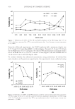
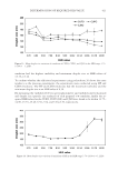
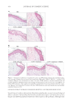
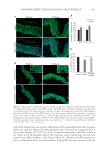
JOURNAL OF COSMETIC SCIENCE 386 occurring during heme catabolism drugs etc.). Acquired forms of hypermelanosis can have several different causes (e.g., metabolic or endocrine disorders, defi ciencies, cutaneous injuries, infl ammatory dermatoses, and systemic and neurological diseases) (2,3). Melasma is the most frequent form of acquired hypermelanosis, occurring most commonly on the face and also in extrafacial areas (4), and is characterized by an increased deposition of melanin (5). Various epidemiological studies estimated the prevalence of melasma at 1% in the general population and 9–50% in higher risk populations (6,7). Morphologically, melasma presents as symmetric reticulated hyperpigmented patches with irregular bor- ders on the centrofacial region, malar cheeks, mandible, and rarely upper chest and ex- tremities (4). Melasma, rather than a rigid linear epidermal problem, is now considered a heterogeneous pathology derived from a complex interplay among melanocytes, kerati- nocytes, dermal fi broblasts, and vascular endothelial cells (8). Melanin is produced by melanocytes in the basal layer of the epidermis from the amino acid tyrosine, within organ- elles known as melanosomes, through a reaction catalyzed by the tyrosinase enzyme. Melanocytes then export mature melanosomes to nearby keratinocytes through their dendrites to induce pigmentation (9). Immunohistochemical studies on skin biopsies confi rmed a signifi cant increase in melanin in melasma but no quantitative increase in melanocytes in the hyperpigmented areas of skin that, however, resulted in larger and very prominent dendrites (10). The factors that can cause an increase in melanin concentra- tion are numerous and include, among others, hormonal and genetic factors, high exposure to ultraviolet rays, darker skin types, some drugs, infections, or infl ammatory processes (6,7). However, the pathogenesis of melasma is not yet fully understood. Increased ex- pression of the stem cell factor in the dermis and of the tyrosine kinase receptor c-kit in the epidermis has been suggested to play an important role in the mechanism of hyper- pigmentation in melasma (11). A relevant role in melasma has also been suggested for increased microcirculation, triggered by a signifi cantly increased level of the vascular endothelial growth factor (VEGF), a major angiogenic factor of the skin constitutively produced by keratinocytes and whose receptors are expressed both in melanocytes and vascular endothelial cells (12,13). The VEGF could have a direct infl uence on melanocyte behavior and melanogenesis through its receptors. Interestingly, the VEGF is known to stimulate the arachidonic acid release and the phosphorylation and activation of the cyto- solic phospholipase A2 (14). It is possible that the resulting metabolites from this path- way affect melanogenesis as well. T he result of increased melanin concentration is an uneven skin tone. Although common, the management of this disorder remains challenging, given the incomplete understanding of its pathogenesis, chronicity, and recurrence rates. Moreover, melasma treatment is quite challenging because of the presence of melanin deposits at varying depths in the epidermis and dermis (15), with dermal and mixed melasma (a combination of the epider- mal and dermal types) having the worse prognosis because many topical therapies are not able to target dermal melanophages (16). Several methods and strategies have been re- ported so far in the literature to address skin hyperpigmentation. The goal of melasma treatment is to decrease melanin production and increase its elimination. In addition to traditional treatments, there are also new p romising strategies, including oral, topical, and procedural therapies (5). Among them, the chemical peeling, also called “chemical resurfacing,” that consists in the application of one or more substances, in immediate or delayed sequence, which causes a chemical ablation of defi ned skin layers. This treatment leads to a uniform and taut skin, through the regeneration and repair mechanisms of the

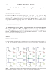
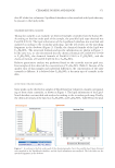
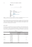
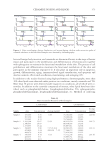
A DEPIGMENTATION TOPICAL TREATMENT PROGRAM ENHANCES A PREVIOUS CHEMICAL PEELING OF THE FACE 387 epidermis and dermis stimulated by a controlled infl ammatory reaction, with the synthe- sis of new c ollagen and a more evenly distributed melanin (15,17). Chemical peels are effective in improving skin tone and reducing melanin concentration. However, outpatient chemical peels can be combined with other home-based treatments to provide a synergis- tic approach and optimize clinical outcomes, enhance patients’ satisfaction, and allow clini- cians to tailor the treatment to individual patient needs and conditions (15,18). In particular, Rendon et al. (15) reviewed several chemical peel protocols applied to melasma, including those based on glycolic acid and salicylic acid, and concluded that a maintenance therapy is needed when peeling is used for melasma, and that chemical peels may be most effective when used in combination with medical therapy or other procedures possibly because peels remove melanin, although other treatments inhibit melanocytes or melanogenesis. C hemical peeling treatments can be classifi ed into three categories: superfi cial peels, which exfoliate the epidermal layers without going beyond the basal layer medium depth peels, which reach to the upper reticular dermis deep peels, which penetrate the lower reticular dermis (18). The depth of peeling, and thus the degree of its therapeutic effects, is affected by different factors, including the properties of the chemical agents used (e.g., concentration and pH), the application technique, and the skin condition and sensitivity. O nce a chemical exfoliant has exhausted its thinning action on epidermal structures, the depigmentation process does not continue further and can be considered stabilized. How- ever, its benefi cial action could be potentiated by subsequent dermocosmetic treatments. Here, we report the results of a monocentric, prospective, noncontrolled study carried out in female subjects submitted to chemical peeling within 7 d before the study inclusion and aimed at the evaluation of the effects of a 30-d topical, depigmenting dermocosmetic treatment program on facial hyperpigmentation, pigment uniformity, and skin texture. M ETHODS S TUDY DESIGN T his was a monocentric, prospective, noncontrolled study carried out between December 2018 and February 2019 (fi rst s ubject enrolled and last subject completed) in Milan, Italy, aimed at the evaluation of the effi cacy, safety, ease of use, pleasantness, and tolerability of a depigmenting topical dermocosmetic treatment program in the reduction of facial hyper- pigmentation, measured as dark spot size reduction, in subjects submitted to chemical peeling within 7 d before the study inclusion. T he protocol, consent form, information sheet, and any written information to be provided to study participants were submitted to an independent ethics committee, and a copy of the written approval was obtained. The study was conducted in accordance with the Italian regulations and requirements. I NCLUSION CRITERIA T he study inclusion criteria were signed informed consent, ≥18 years of age, a good general health condition, and superfi cial depigmenting chemical peeling treatment within 7 d before the study enrollment with the Defi nisse™ Peel Program (Mastelli Srl, Sanremo, Italy).
Purchased for the exclusive use of nofirst nolast (unknown)
From: SCC Media Library & Resource Center (library.scconline.org)