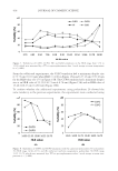
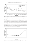
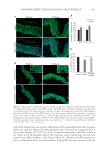
JOURNAL OF COSMETIC SCIENCE 428 1 × 2 min in eosin, 1 × 2 min in 95% ethanol, 2 × 2 min in 100% ethanol, and 2 × 2 min in xylene. Coverslips were mounted on glass slides using Eukitt® (xylene based) as mount- ing media (O. Kindler, Germany) and dried overnight. IMMUNOHISTOLOGICAL FLUORESCENCE Human sk in samples were obtained from pl astic surgery of healthy females who had given written informed consent. After removal of subcutaneous fat, the tissue was used to obtain 6 mm punch biopsies that were incubated with the patchouli extract (or its pla- cebo) at 1% for 48 h. Biopsies were then fi xed in formaldehyde and processed in an auto- mated Shandon Hypercenter XP (Shandon Ltd., Runcor, United Kingdom) for paraffi n embedding. Sections of 4 μm thickness were cut with a microtome (Shandon) and col- lected on polylysine-coated glass slides (Menzel Gläser, Braunschweig, Germany) for im- munohistochemistry. Heat and pepsin enzymatic antigen retrieval was performed before incubation with CB2 receptor and β-endorphin antibodies. Heat antigen retrieval was performed before incubation with IL1R1 primary antibody. Pepsin enzymatic antigen retrieval was performed before incubation with TRPV1 primary antibody. No antigen retrieval was performed before incubation with IL6ST primary antibody. After incubation with the secondary antibody, cell nuclei were stained with 4′,6-diamidino-2-phenylindole (DAPI). Skin sections were viewed under a microscope (Axiovert 200M, Carl Zeiss, Oberkochen, Germany) and photographed with a CCD camera (EXI blue, Qimaging, Surrey, BC). SUBJECT To demonstrate the potential effect of patch ouli extract on sensitive skin, a stinging test was performed as the reference test for sensitive skin. However, other parameters known to be altered on sensitive skin, like skin barrier function, were evaluated on these 26 volunteers on the forearm at the same time as the stinging test (data not shown). One month before the beginning of the clinical test, panels of subjects who are sensitive to 0.01% of capsa- icin diluted in 10% ethanol (which cause sensory irritation) and not to 10% ethanol, applying on nasolabial fold during 10 s, were preselected by a trained expert. Just after the exposure, the subject recorded the sensations of stinging each minute during 10 min on a 0- to 4-interval scales (0 = no sensation, 1 = slight stinging, 2 = moderate stinging, 3 = intense stinging, and 4 = very intense stinging). At the end of the 10 min, the 11 values recorded by the subject were summed up to a fi nal score. If this score was superior to 10, the volunteer was declared like having sensitive skin. Only volunteers having a score superior to 10 were identifi ed as having sensitive skin and were enrolled in the study. At the beginning, 26 volunteers were enrolled in the study and were divided in two groups of 12 volunteers homogenous in age and gender to be sure that these two parameters did not infl uence the results. However, during the test period, six volunteers had to be excluded of the stinging test results, because of big variation in their stinging sensation compared to other trials done at the same period. For fi ve of these volunteers, the variation was explained by a bad cold, and for the last one, no explanation was found. Thus, the stinging result was performed on 20 volunteers, divided in two groups of 10 volunteers homogenous in age and gender.

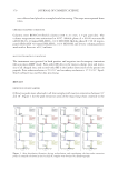
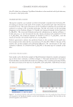
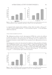
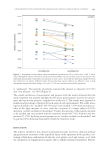
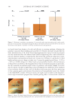
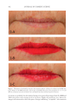
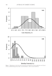
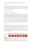
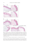
SOOTHING EFFECT OF POGOSTEMON CABLIN EXTRACT 429 FACE CAPSAICIN–INDUCED STINGING TEST For thi s clinical study, 20 volunteers were enrolled after having read the information re- lated to the study and signed the informed consent form. All volunteers had sensitive skin and were divided in two groups of 10, homogeneous in age and gender. For 28 d, on all the faces, one group applied a placebo cream and one group applied a cream containing the patchouli extract at 1%. The study was performed in double blind. The stinging test was carried out on the fi rst day of the study (D0), before cream application and 28 d after cream applications (D28). The test consisted in applying 0.01% of capsaicin diluted in 10% ethanol to one nasolabial fold and 10% ethanol to the other during 30 s. Just after the exposure, the subjects recorded the sensations of stinging each minute during 10 min on a 0- to 4-interval scales (0 = no sensation, 1 = slight stinging, 2 = moderate stinging, 3 = intense stinging, and 4 = very intense stinging). At the end, the 11 scores were summed to give a score of skin sensitivity. STATISTICAL ANALYSIS All experiments have be en repeated at least twice with cells and skin coming from differ- ent plastic surgery, and the quantifi cation of the fl uorescence was performed by measuring the fl uorescent intensity of the staining, which was normalized by the epidermis or cell area. Statistical analyses were performed using Excel 365 (Microsoft, Redmond, WA). Difference between two means was performed with Student’s t-test. A p-value d 0.05 was considered statistically signifi cant (*), p-value d 0.01 as very signifi cant (**), and p-value d 0.005 as highly signifi cant (***). For the clinical test, statistical analysis was performed using JMP® 14 (SAS Inc, Cary, NC) software. The homogeneity of the gender between the two groups was performed using the χ2 test, and the homogeneity of the age was confi rmed using the Mann–Whitney test. Concerning the statistical analysis of stinging values, the scores, which were used as a basis, concerned the difference (D28–D0) for the patchouli extract and placebo group using Wilcoxon test (after verifying the data followed a normal distribution or not with the Shapiro–Wilk test). RESULTS THE CB2 RECEPTOR ACTIVATION SHOWS RESISTANC E TO UV- INDUCED SKIN INFLAMMATION Because of the potential role of cannabinoids in hu man skin infl ammation, we investigated whether the antagonist AM630 would exhibit an amplifi ed susceptibility to UVB- induced infl ammation in human skin biopsies. Histologic analysis indicated that UVB- treated human skin showed a pronounced infl ammation with the emergence of structural damages and sunburn cells (Figure 1A). Exposure to AM630 (3 h) before UVB irradiation showed a very damaged structure with an important epidermal erosion, whereas the epi- dermis exposed to AM1241 (3 h) was far more resistant to UVB-induced infl ammation (Figure 1A). In a second time, skin was treated with the patchouli extract during 48 h after UVB irradiation. The biopsies were treated or not during 3 h with AM630 prior UVB irradiation. The patchouli extract preserved the skin from UVB, compared with the placebo condition (Figure 1B). The antagonist AM630 was able to partially block the protective effect of the patchouli extract on skin preservation.
Purchased for the exclusive use of nofirst nolast (unknown)
From: SCC Media Library & Resource Center (library.scconline.org)