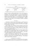
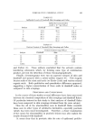
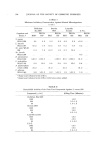
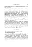
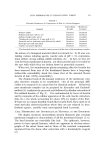
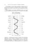
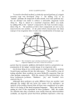
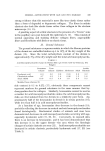
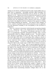
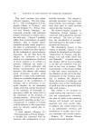
CELL MEMBRANES IN KERATINIZED TISSUE 511 TABLE I Chemical Resistance of Components of Hair to Various Reagents Intracellular Membrane- Reagent Keratin Complex a Sodium sulfide Sodium hydroxide (pH 11-12) Sodium thioglycolate, pH 11 Thioglycolic acid plus 10 N urea (pH 6-11) Peracetic and/or performic acids followed by alkali Hydrogen peroxide and alkali Tryptic and peptic digestion Dissolves Resistant Dissolves Resistant Dissolves Insoluble Dissolves Resistant Dissolves Resistant Dissolves Resistant Resistant Dissolves The chemical behavior of medulla (when present) is like that of the membrane-complex. the nature of a biological material which is resistant to: 10 M urea con- taining various reducing agents, caustic soda of pH 12, cuprammo- nium sulfate, strong sodium sulfide solutions, etc. In fact, no true sol- vent for these membranes is known yet their protein basis is revealed by the ease with which they are broken down by proteolyic enzymes. When wet, the membraneous ghosts remaining after the keratin has been removed from any of the keratinized tissues have a long-range rubber-like extensibility about five times that of the oriented fibrous keratin of hair (100% extensibility). The chemical basis of the unusual resistance of the membrane com- plex therefore remains to be established. One of the principal diffi- culties is to separate it in a pure form for chemical analysis. Relatively pure membrane-complex can be prepared by Alexander and Earlands' method (4) (oxidation by peracetic acid followed by alkaline extraction of the oxidized keratin, cf. Fig. 2). It is contaminated by traces of keratin and by remnants of the cellular apparatus of the cells (nuclei, mitochon- dria, etc.), the whole accounting for about 10% by weight of the fiber. If it proves to contain disulfide bonds like keratin itself, these must be in some sterically sheltered position where they are not reduced by kera- tinolytic agents possibly some unrecognized bond is present. CELL MEMBRANES AND INTERCELLULAR MATERIAL IN THE EPIDERMIS The duplex structure (intracellular keratin filaments plus resistant membrane-complex) is characteristic of all the keratinized tissues (5, 6). The hard keratins are essentially like hair. The epidermis, however, proves to have special features, which are apparent in electron micro- graphs of the intact tissue and in those of the resistant components separated from the tissue after extraction with a keratino!ytic reagent (7).

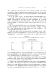
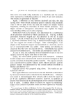
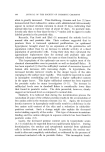
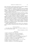
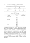
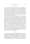
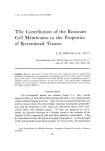
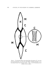
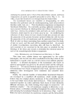
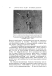
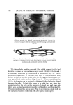
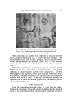
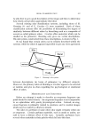
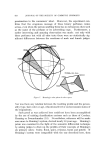
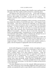
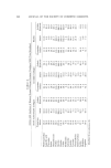
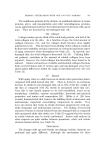
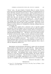
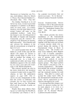
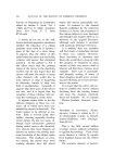
512 JOURNAL OF THE SOCIETY OF COSMETIC CHEMISTS Figure 3. An electron micrograph of a section of the stratu•n corncure (human) showing the limited development of adhesive patches (at arrows). Compare with the continuous layer of bonding material in hair (Fig. 2) Figure 4. Drawing illustrating the patchy nature (P) of the intercellular adhesive (C) substance in the upper layers of the stratum corneum (M-cell •ne•nbranes, K-keratinized contents of cells) The intercellular bonding material (also called cement) in the hard keratins is a more or less continuous layer about 400-600 A thick, and it is essentially unaltered by the removal of the keratin (Fig. 2). In the keratinized epidermis, in contrast, this layer is discontinuous, being limited to discrete patches which unite the two opposed membranes over only part of their entire surfaces (Figs. 3 and 4). These rounded patches (1-2 u) in diameter can be pictured as flattened balls of adhesive slipped between the cells. In fact, this seems to be what they are, since they originate within the cells as the contents of closed sacs and open onto their faces, as has been shown recently by Matoltsy and Parrakal (8). These rounded patches can be seen (Fig. 4) as numerous small studs on the surfaces of isolated epidermal cells after special staining (9).
Purchased for the exclusive use of nofirst nolast (unknown)
From: SCC Media Library & Resource Center (library.scconline.org)