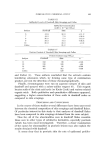
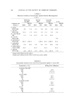
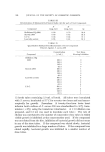
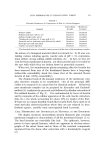
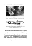
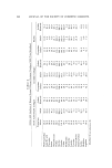
DERMAL ALTERATIONS WITH AGE AND SUN DAMAGE 529 strong evidence that this material is more like true elastic tissue rather than a form of degraded or degenerate collagen. The fibers in actinic elastosis also look like elastic tissue rather than collagen in the electron microscope (14, 15). A puzzling aspect of actinic elastosis is the presence of a "Grenz" zone in the papillary area just beneath the epidermis (8, 12). This consists of normal appearing and staining delicate collagen fibers, argyrophilic fibers and fibroblasts with little or no elastic tissue. II. Ground Substance The ground substance or aqueous matrix in which the fibrous proteins of the dermis are embedded makes up 5 to 10% of the dry weight of the dermis (16). Since the total carbohydrate content of the dermis is approximately 1% of the dry weight and the total acid mucopolysaccha- TABLE I Acid Mucopolysaccharide (Amps) in Human Skin (um Uronic Acid via Orcinol/g. Dry Weight) Premature Term Actinic Infants Infants Children Adolescent Adult Elastosis Hyaluronic acid 6.0 5.0 3.8 0.61 0.9 3.2 Chondroitin sulfate 4.7 4.2 1.0 0.96 1.3 1.6 Total AMPS 10.9 9.5 4.9 1.8 2.4 5.2 Modified from reference 23. ride content is 0.1 to 0.2% (17), obviously these components cannot represent markers for ground substance in the same manner that hy- droxyproline does for collagen. Similarly, hexosamine cannot be used as a marker for acid mucopolysaccharides unless the acid mucopolysaccha- rides are first isolated from the dermis in a relatively pure form. Ap- proximately half of the hexosamine in dermis is in serum proteins (18), while less than half is in acid mucopolysaccharides. As a function of age, hexosamine does decrease in the dermis (19), probably reflecting the decrease in neutral and acid mucopolysaccharides found histologically. Biochemical studies of animals and man have demonstrated decreases in acid mucopolysaccharides occurring with age, especially hyaluronic acid (17, 20, 21). Conversely, in exposed skin, there is an increase in hexosamine, and it has been demonstrated that this increase is in the upper dermis--the area where the histologic changes of actinic elastosis are seen (22). Acid mucopolysaccharides are increased in actinic elastosis, particularly hyaluronic acid (17, 23) (see Table I).

530 JOURNAL OF THE SOCIETY OF COSMETIC CHEMISTS
Purchased for the exclusive use of nofirst nolast (unknown)
From: SCC Media Library & Resource Center (library.scconline.org)