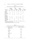
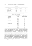
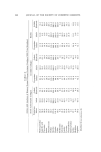
DERMAL ALTERATIONS WITH AGE AND SUN DAMAGE 5B1 The non fibrous proteins in the dermis, an undefined mixture of serum proteins, glyco- and mucoproteins and other noncollagenous proteins, can be approximated and have been demonstrated to decrease with age in man. They are increased in sun-damaged skin (16). III. Collagen Collagen makes up one-third of the total body protein, and half of the total collagen is in the skin. As a function of age, the total amount of collagen increases (16), and the collagen itself becomes more highly polymerized (24). This increased cross-linking of the collagen results in its decreased solubility and may represent an extremely important aspect of aging connective tissue throughout the body (25). In exposed, sun- damaged skin, the total collagen is decreased (16, 26). Utilizing differ- ent patients, variations in the amount of soluble collagen have been reported however, the total collagen has invariably been found to be reduced. Amino acid analyses of soluble and insoluble collagen fractions from covered human skin of various ages and sun-damaged areas have shown minor differences within the range of experimental error (26) (see Table II). IV. Elastin With aging, there is a slight increase in elastin when premature skin is compared with adult human skin (26). There is, however, an enormous increase in elastin in sun-damaged skin, up to 13% of the dry weight of the skin as compared with 2% elastin in unexposed adult skin (27). That this is true elastin appears to be well established, based on its morphology, solubility, enzyme susceptibility, tinctorial and physical properties, and amino acid composition (28). Miller et al. (29) have recently demonstrated that lysine is the buildiug block of desmosine and isodesmosine, important cross-linking components in elastin. They have also shown that lysine in elastin decreases progressively with age, while desmosine and isodesmosine increase. The finding of increased amounts of lysine in elastin isolated from sun-damaged skin as compared with unexposed adult skin elastin (see Table II) suggests that the elastin in actinic elastosis may be newly synthesized. Ami•o acid analyses in other respects are quite similar between premature, v.dult, and actinic elastosis dermal elastin (see Table II). DISCUSSION The changes with age and chronic sun damage (actinic elastosis) are profound and quite different. Although the mechanism for these

532 JOURNAL OF THE SOCIETY OF COSMETIC CHEMISTS changes is not known, hypotheses based on some recent studies may ex- plain what is happening. Sub cellular particles called lysosomes (30) have been demonstrated in fibroblasts and are known to contain col- lagenase (31, 32). Elastase has not been reported in such particles, and its only known mammalian source is the pancreas (13). The lysosomes are labilized by heat and ultraviolet light below 3100 A (33, 34). One m.ight postulate that the labilization of these particles with release of collage- nase leads to the digestion and thus reduction of the amount of collagen in the dermis following sun exposure. The extent of these changes, of course, would be dependent upon natural protection such as pigmenta- tion and individual variability of the susceptibility of the lysosomes to labilization. There are enzymes in lysosomes which degrade mucopolysaccharides (30, 35). Hyaluronie acid is also depolymerized by ultraviolet light below 3100 A (36, 37). The increase of hyaluronic acid may be a func- tion of enzymatically degraded mucopolysaccharides being recycled metabolically while the collagen sub units are not. The lack of elasto- lyric enzymes in lysosomes as well as the lack of any direct effect or ultraviolet light (2900-3200 A) in degrading either collagen or elastin in vitro (38) could explain the increase in elastic tissue found in this disorder as new connective tissue, collagen and elastin, is synthesized to replace the digested collagen. It must be emphasized that it is not known if ultraviolet light in the crucial range of 2900-3100 A labilizes lysosomes. The presence of the "Grenz" zone of normal appearing connective tissue associated with argyrophilic fibers just beneath the epidermis and the depth of the elastosis below the level of penetration of ultraviolet light from natural sunlight may be clarified by some recent studies in amphibia. Using 3H proline Hay and Revel (39) have shown concentra- tion of the label at the epidermal-lamellar junction, suggesting that newly formed collagen of the dermis is first deposited at this site. In the lam- prey, the collagen fibrils nearest the epidermis are small (as in man), but in the deeper layers beneath the epidermis they become larger, presuma- bly representing older more mature fibers (40). Therefore, it appears that the dermis and epidermis grow in opposite directions from each other, that is, the epidermis grows outward and the dermis grows inward, possibly pushing newly synthesized elastotic fibers along with it. Such growth of the dermis would satisfactorily explain a number of other observations such as: the fine delicate fibers in the papillary layer and the thicker coarser fibers in the reticular layer of unexposed skin the argyrophilic fibers representing immature collagen fibers (41) in the
Purchased for the exclusive use of nofirst nolast (unknown)
From: SCC Media Library & Resource Center (library.scconline.org)