J. Soc. Cosmetic Chemists, 16, 527-535(1965) Dermal Connective Tissue Alterations with Age and Chronic Sun Damage J. GRAHAM SMITH, JR., M.D., and G. ROLLAND FINLAYSON, M.D.t Presented December 2, 1964, New York City Synopsis--The changes in human Caucasian skin commonly believed to be due to aging are primarily the effects of prolonged repeated damage to the skin from the sun. Covered aged skin shows marked differences histochemically and biochemically from exposed aged skin. With aging there is a decrease of non fibrous protein and of soluble collagen, although the total collagen increases. The total aci• mucopolysaccharides decrease, especially hyaluronic acid. In chronically sun-damaged skin (actinic elastosis) there is little change in the amount of extractable soluble collagen. The insoluble collagen content is reduced to one-third of that of control skin, and there is an increase in an elastin-like protein. Total acid mucopolysac- charides increase in actinic elastosis, especially hyaluronic acid. INTRODUCTION Exposure of susceptible Caucasians to the elements, especially sun- light, is more important than age in producing the clinical changes of actinic elastosis--wrinkling, loss of elasticity, and histologic changes in the dermis. This has been appreciated by careful investigators of the problem for over 75 years (1), and confirmatory studies have been re- ported by many investigators (2-5). Indeed, Benjamin Franklin (6) in * Supported in part by National Institutes of Health Grants AM 05812, AM 07583 and ST1 AM 5335. t Division of Dermatology, Department of Medicine and the Center for the Study of Aging, Duke University Medical Center, Durham, N. C. 527

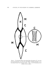
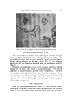
528 JOURNAL OF THE SOCIETY OF COSMETIC CHEMISTS 1745 may have been alluding to this when he stated that" . . . covering all above with a basket, and regarding only what is below the girdle (waist), it is impossible of two women to tell an old one from a young one" since the changes of actinic elastosis which are so commonly inter- preted as changes of age are not observed clinically or histologieally in unexposed areas such as the lower abdomen and buttocks. Pigmentation of the skin represents a natural protective mechanism from these changes, and it is a common clinical experience to have difficulty judging the age of Orientals and Negroes, both of whom show much less severe changes in the exposed skin with age. This paper will review the histologie and biochemical alterations occurring in the dermis with age and chronic sun damage (actinic elastosis). AGING AND SUN DAMAGE I. Histologic and Electron Microscopic Changes In reviewing the literature concerning cutaneous aging changes, it is sometimes difficult to be sure that investigators have appreciated the striking differences which may occur in exposed skin as compared with covered skin. With age, few histochemical changes are observable in the human dermis. Small decreases in neutral and acid mucopolysaccha- rides are associated with thickening and coarsening of the collagenous fibers (7). Few dramatic changes are seen in the elastic fibers, although there appears to be a slight increase in the skin of adolescents and adults as compared to premature infants. Sun-exposed skin from Caucasians shows dramatic changes indeed, the changes are seen to a lesser extent in exposed skin from Negroes (3). Using the periodic acid-Schiff stain after diastase digestion for neutral mucopolysaccharides and the Mowry colloidal iron or alcian blue stains for acid mucopolysaccharides, there are increases in neutral and acid mucopolysaccharides (8, 9). In the upper portions of the dermis, there is marked basophilia with toluidine blue and atypical staining with the van Gieson, aniline blue, phosphotungstic acid hematoxylin (8), and luxol fast blue stains (10). Elastic tissue stains such as orcein, Verhoeff's, and aide- hyde fuchsin stain the fibers heavily in the upper dermis (11, 12). These fibers which stain like elastic tissue are digested by elastase but not by collagenase or crystalline trypsin (11). If these fibers were collagen, they should be digested by collagenase, and if they were degraded col- lagen, they should be digested by both collagenase and trypsin (13). The lack of susceptibility of these fibers to either collagenase or trypsin is
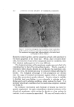
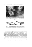
Purchased for the exclusive use of nofirst nolast (unknown)
From: SCC Media Library & Resource Center (library.scconline.org)