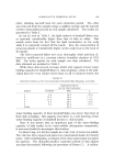
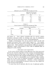
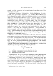
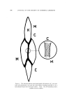
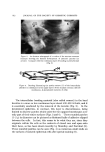
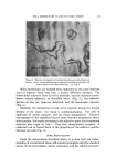
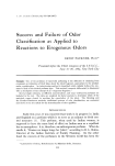
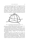
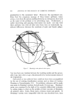
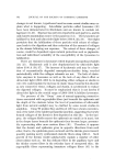
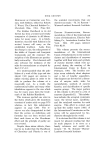
CELL MEMBRANES IN KERATINIZED TISSUE 513 Figure 5. Electron micrograph of resistant membranes isolated from epi- dermis. Note: the membranes have separated, and the intercellular ad- hesive patches have been dissolved. Cf. Fig. 3 When membranes are isolated from epidermis by the same methods used to separate them from hair, a further difference emerges. The intercellular adhesive layer in hair is insoluble, and the separated mem- branes remain adherent, as described above (Fig. 2) the adhesive patches in skin are, however, dissolved, and the membranes separate (Fig. 5). Similarly, the membranes in hair never separate during the normal lifetime of the tissue the tissue is nondesquamating. The cells of epidermis of course separate, and the tissue desquamates. Electron micrographs of the superficial layers show that the membranes them- selves persist, the breaks occurring in the adhesive spots (the keratinized contents also begin to fray). Thus this characteristic property of epidermis can be traced down to the properties of the adhesive patches between the cells (Fig. 4). SOME REFLECTIONS From the observations described above, it is clear that our under- standing of a keratinized tissue will remain incomplete until the chemical nature of the intercellular cement substances and the altered cell mem-

514 JOURNAL OF THE SOCIETY OF COSMETIC CHEMISTS branes are known. The first step is the separation of each of the com- ponents in a pure form and in adequate amounts. An adequate analysis should answer the following questions: (a) What chemical events occur during keratinization to change the labile phospholipid-protein complex of the living cell membrane into the extraordinarily resistant substance found in the hardened tissue ? (b) Similarly, what is the chemical composition of the resistant inter- cellular bonding substance which unites the altered membranes ? (c) What is the chemical difference between the intercellular material in hair and that in epidermis, which causes one to be permanent and the other to disintegrate ? These separations have been attempted and have not proved entirely satisfactory up to date they could probably be perfected were effective use to be made of electron microscopy to control the purity of the prepa- rations. Hopefully, we can look forward to having answers to the above questions before too long. (Received August 20, 1964) REFERENCES (1) Astbury, W. T., and Woods, H. J., Phil. Trans. Royal Soc. (London), A232, 33 (1933). (2) Birbeck, M. S.C., and Mercer, E. H., J. Biophys. Biochem. Cytol., 3,203,215, 227 (1957). (3) Mercer, E. H., Keratin and Keratinization, Pergamon Press, Oxford and New York (1961). (4) Alexander, P., and Earland, C., Nature, 166, 396 (1950). (5) Mercer, E. H., Textile Res. J., 23,388 (1953). (6) Idem, Nature, 172, 164 (1953). (7) Brody, I., J. Ultrastructure Res., 2,482 (1959) 3, 84 (1959) 4, 264 (1960). (8) Matoltsy, A. G., and Parrakal, P. F., J. Cell Biol., 24, 297 (1965). (9) Kligman, A.M., The Biology of the Stratum Corneum, in Montagna, W., and Lobitz, W. C., The Epidermis, Academic Press, N.Y. (1964).
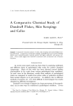
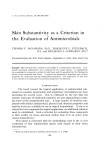
Purchased for the exclusive use of nofirst nolast (unknown)
From: SCC Media Library & Resource Center (library.scconline.org)