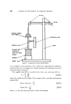
486 JOURNAL OF THE SOCIETY OF COSMETIC CHEMISTS methanol has been found (102) to reduce this interference, but the pre- paration of a blank extract of an adjacent area of the layer was still necessary for full correction other errors were introduced by incomplete elution from the absorbent. Dimethoate and its oxygen analogue have been determined in vegetable tissue by Steller and Curry (81) who digested the eluted pesticides with a nitric-perchloric acid mixture and evaluated the liberated phosphorus by a molybdenum blue spectrophotometric procedure. Dinoseb has been determined spectrophotometrically as its sodium salt after a clean-up on wedge-layer plates (36). Infra-red spectrometry has also been used for the quantitative deter- mination of organo-phosphorus pesticides (34) as well as simultaneously providing conclusive evidence of the identity of the separated pesticide. Gas-liquid chromatography provides a very convenient method for the determination and further clean-up of the eluted pesticide spots. The use of electron-capture detection for both organo-chlorine and organo- phosphorus compounds (41,103) gives very great sensitivity. The use of a silver nitrate-uv irradiation procedure for the visualization of the chromatogram leaves the bulk of the pesticide unaffected in each spot since only the surface of the layer comes under the influence of the irradiation. Thus elution of the pesticide from the treated chromato- plate with dichloromethane gives ample material for an approximately quantitative evaluation. If insufficient material is present to give a visible spot with the visualization reagent used, then elution of that area of the layer suspected of containing the pesticide may give sufficient material for further examination by gas-liquid chromatography. Biological assay, using Drosophila melanogaster, has been used by Salo et al (79) to determine some organo-chlorine and organo-phosphorus pesticides separated on silica gel containing built-in fluorescein as indicator. Marco and Jaworski (104) utilized C •4- labelled Colep for the determination of residues of the pesticide and its various metabolites by radiometry. A suitable scanner for thin-layer chromatography has been described by Wilde (105). A useful description of the sequence of techniques involved in the routine quantitative analysis of materials which have been separated by thin-layer chromatography has been given by Millett, Moore and Seaman (106). Application of the samples, removal of the layer material bearing the located compound, and the elution of the latter from the absorbent are detailed the accuracy and precision (1-2%) of the techniques are also discussed.

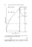
THIN-LAYER CHROMATOGRAPHIC TECHNIQUES IN RESIDUE ANALYSIS 487 The second class of methods used for the quantitative evaluation of thin-layer chromatograms follows more closely those techniques which have been well established in the field of paper chromatography (107). Spot density measurements have been used by Privett and Blank (108) in the determination of component triglycerides. The areas under the densitometer curves were found to be directly proportional to the amount of sample applied for the saturated triglycerides and glyceryl residues of the unsaturated glycerides, spots being located by charring with 50% sulphuric acid. However, the area given varied with the type of structure of the compound. Squibb (109) made densitometric measurements on amino-acids located with ninhydrin and found a coefficient of variation of q- 6.90/0 . Reflectance densitometry as used for the paper chromato- graphic determination of herbicides (83) has been found to be less success- ful with thin-layer chromatograms of similar compounds, largely owing to the fragile nature of the layer surface and the difficulty in obtaining a sufficiently clean and uniform background w•,•h the silver nitrate reagent used. A review of photometric methods has been given by Klaus (110). Methods based upon the measurement of spot area avoid the difficulties associated with elution of the material from the absorbent and the possi- bilities of further pollution of the purified sample. Seher (111) simul- taneously chromatographed samples of the unknown and a series of standards applied to the chromatoplate in equal volumes of solution. After rendering the developed chromatogram visible, the areas occupied by the standard sample spots were determined and plotted against the corresponding weight of material. Reference of the area of the unknown sample spot to this curve gave a measure of the material present. Purdy and Truter (112) showed that the square root of the area of the spot is a linear function of the logarithm of the weight of the material it contains. Statistical evaluation (113) showed that this relationship was preferable to those of area against logarithm of the weight or logarithm of the area against logarithm of the weight. Planimetric means were used by Aurenge et al (114) to determine spot areas graphs of area squared against weight of material were linear. They also traced the chromatograms, cut out the traced spots and weighed them in order to determine the area of the spot more accurately, the weight per cm • of the tracing paper being known. (Received: 14th July 1965)
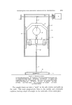
Purchased for the exclusive use of nofirst nolast (unknown)
From: SCC Media Library & Resource Center (library.scconline.org)