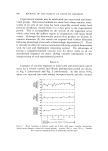
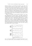
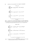
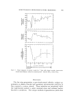
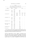
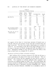
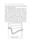
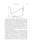
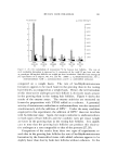
J. Soc. cromer. Chem., 21, 845-852 (Dec. 9, 1970) Direct Observation and Ouantitation of Subcutaneous Microcirculatory Responses to Exogenous Agents GENE J. YOUKII.IS, Ph.D.,* ROBERT C. MARTZ, M.D.,* and PATRICK D. HARRIS, Ph.D.* Presented December 2, 1969, New York City Synopsis--Direct observation of subcutaneons minute vessels found in the wing of the un- anesthetized small brown bat (Myotis luci[ugus) is accomplished by a compound MICRO- SCOPE-closed circuit TELEVISION system. This technique represents an excellent method to evaluate directly in an unancslhctized mammal the SUBCUTANEOUS MICROVASCU- I•AR REqPONSES to systemically injected, locally injected, and topically applied exogenous agehis. S•nall artery (35--45 /•) and small vein (75-85 /•) diameters are measured at gO-second intervals by marking the position of vessel walls on a procalibrated television monitor screen. Small vein vasomotion (cycles of alternating constriction and relaxation) is counted over 1-minnie interxals. Both control and experimental animals are divided into intact vascular innervalcd and vascular dcnervated groups to evaluate vaScnlar responses with and without central neuronal control. Control animals are injected intraperitoneally with normal saline while experimental animals are injecled similarl) with various exogenous agents. The bat wing preparation was originally utilized by Nicoll anti Webb (1) for structural and functional studies of minute blood and * Present address: Depart•nent of Pathology and Toxicology, G. 1). Searle and Co., P.O. Box 5110, Chicago, Ill. 60680. Investigation performed at Indiana University Medical Center, Indianapolis, Ind. 46202. ?Deparlment of Toxicology, Indiana University Medical Center, 1100 West Michigan Street, Indianapolis, hid. 46202. • Del)artment of Physiology, University of Missouri School of Medicine, Columbia, Mo. 6521) l. 845

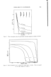
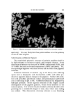
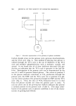
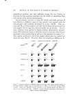
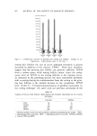
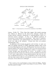
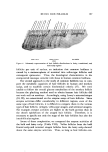
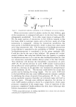
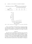
846 JOURNAL OF THE SOCIETY OF COSMETIC CHEMISTS lymphatic vessels. The very thin wing membrane provides an area of a peripheral vascular bed easily viewed directly without the need for anes- thesia and further surgical manipulations. At present very little experimentation has been accomplished with this preparation in which chemical agents have been applied to the intact epidermal surface of the wing. Wiedeman (2) studied vascular reactivity following denervation of subcutaneous areas of the bat wing in an effort to determine the contribution of neural and humoral factors to adjust- ments in vascular beds. She topically applied epinephrine to vessels which had previously been exposed by the stripping of the epidermis of a small area of the wing. The epidermis was removed with a fine forceps in a careful manner so as not to disturb the underlying structures. In addition, Nicoll (3) mentions the direct vascular application of other vasoreactive agents such as norepinephrine, histamine, and acetylcholine which have also been utilized in previous studies. The potential of this preparation, although used mostly in the recent past for microcirculatory studies following systemic administration of exogenous agents, can be extended to studies of topically applied stimuli on the intact epidermis. The purpose of this presentation is to describe the technique as it has been used in recent investigations to yield direct information of microcirculatory system responses to physiologic, pharrna- cologle, and toxicol ogic stimul i. EXPERIMENTAL Procedures The experimental preparation is similar to that described by Nicoll and Webb (1) as modified by Harris et al. (4). The unanesthetized bat is secured on its dorsal surface and both wings are held in an extended po- sition over a glass plate by padded spring clips. The preparation is mounted on the stage of a binocular compound microscope, allowing selection of small arteries (approximately 35-45 v) and small veins (ap- proximately 75-85 v) to be observed. I,ight from a 300-watt zirconium arc lamp is transmitted through the microscopic field, gathered by an ob- jective lens (X21) and divided into two components by a beam splitter. Approximately 90% of the gathered light is passed through a focusing lens (X15) to an Ampex television camera which generates a composite video signal. The video information is analyzed on a 23-in. monitor (total magnification of 1,000x).
Purchased for the exclusive use of nofirst nolast (unknown)
From: SCC Media Library & Resource Center (library.scconline.org)