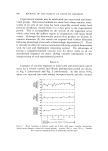
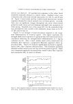
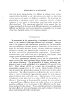
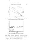
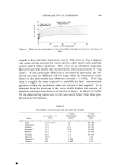
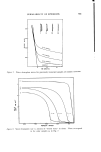
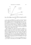
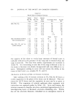
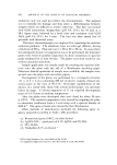
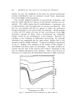
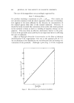
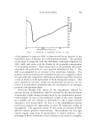
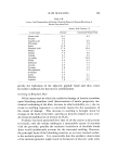
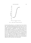
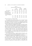
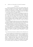
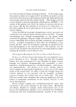
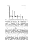
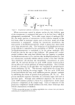
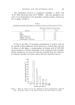
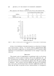
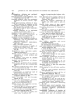
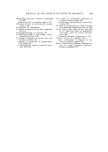
PERMEABILITY OF EPIDERMIS 859 solvents: ethanol, acetone, ether, and a (2:1) chloroform:methanol mixture (2:1, C:M). The solvent was placed only in the receptor half of each diffusion cell water and HTO were placed on the donor side in contact with the stratum comeurn directly. Thus the partition coefficient for water was approximately unity as before. The observed k vvalues indicate that each solvent damages the membrane to a dif- ferent degree with (2:1, C:M) having by far the largest effect. It is no coincidence that (2:1, C:M) is the best lipid solvent of the four, particularly in its capability to extract bound polar lipids and proteolipids as well as neutral lipids. Mechanism of Permeability through Delipidized Tissue T]qe gross appearance of stratum corneum after treatment with (2:1, C:M) is unchanged although the dry weight may decrease by as much as 20% due to lipid extraction. This suggests the formation of submicroscopic interstices or micropores in delipidized tissue and the likelihood of diffusion through them. If this picture is correct, then the resultant permeability should exhibit the following three charac- teristics: (a) The permeability of all molecules should increase mark- edly (b) the permeability of similarly sized polar and nonpolar mole- cules should be equally large since their lipid sorption affinity would now be irrelevant to the major diffusion pathway, i.e., through solvent- filled pores and (c) the activation energy for diffusion should decrease to a value consistent with liquid-state diffusion, i.e., near 4.6 kcal/mole for water. These predictions were tested for stratum comeurn membranes which had been soaked in (2:1, C:M) for 1 hour and then rinsed and soaked in water for 3 days. Arrhenius plots for water diffusion are compared in Fig. 5. At 25øC, the average diffusion constants for water through untreated tissue, delipidized tissue, and through liquid water (self-diffusion) are respectively: 5 X 10 -•ø, 5 X 10 -s, and 2 X 10 -• cm2/sec. Permeability constants for water thus increase approximately 100-fold after 1 hour's soaking the change in permeability after a 5-min soak was very much less. The activation energy has decreased (this may be seen from the slopes of the lines) from 14.3 to 6.3 kcal/mole. As shown in Table I, the permeability constants of small polar and nonpolar alcohols through the delipidized tissue are equally large and independent of their lipid solubility.

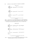
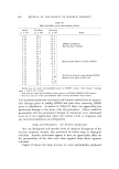
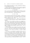
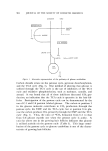
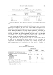
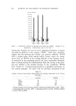
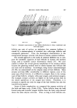
860 JOURNAL OF THE SOCIETY OF COSMETIC CHEMISTS 25'C -7 -8 -9- -•0 • • oo Through dehpldlzed hydrated stratum carneurn - ß E=44 3+ Z e• e Untreatedhydroled •,.....x.....x•_•e ß stratum comeurn •x,-.x,..xo .o x x 5.10 •.20 $.$0 :3.40 •.50 5.60 570 1000 TøK Figure 5. Arrhenius plots for water diffusion Tablc I Pcrmcability of Normal and Dclipidizcd Tissue Propanol Heptanol Normal Dclipidized Normal Dclipidizcd kp X 10 • at 30øC in cm.hr -• 1.7 230 37.6 243 E, in kcal- mole -• 19.6 •---5.0 11 . 0 •-5.0 These data indicate that treatment with (2:1, C:M) for one hour or more results in a fairly porous, nonselective membrane. The low activated ener•oy indicates liquid-like diffusion through water-filled channels in the tissue. The tissue remains a significant water barrier as shown by comparison with the self-diffusion constant of water. This is attributed to the relatively small fractional pore area produced by the solvent treatment (/'po,.e• • 2.5 X 10 -a, as estimated by the ratio of the two tissue diffusion constants). Obviously the individual effective pore diameters must also be quite small. Water and Water-Miscible Liquids Water It is well known that stratum corneum gradually softens and swells in water. As hydration of the tissue proceeds its permeability increases,
Purchased for the exclusive use of nofirst nolast (unknown)
From: SCC Media Library & Resource Center (library.scconline.org)