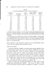
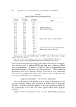
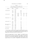
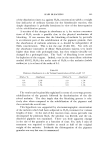
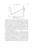
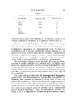
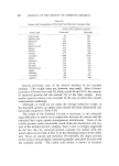
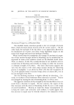
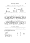
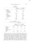
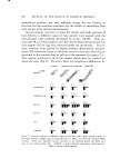
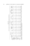
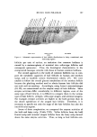
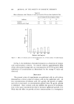
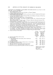
PERMEABILITY OF EPIDERMIS 871 l ipid and non fibrous protein, are arranged roughly parallel to the plane of the membrane. For the membrane to expand and at the same time maintain intercellular coherence as it does, it is necessary either that the helical keratin filaments must individually uncoil and extend or in some way slip relative to each other. It is hard to conceive of a mechanism which is both consistent with the membrane structure and the chemistry of the system that can explain the slipping of filaments. On the other hand, it is known that aqueous surfactant solutions can disaggregate proteins. It is there[ore plausible to suppose that the laurate anion initiates the uncoiling and extension of oz-keratin filaments producing fi-keratin and an expanded membrane. The birefringence of the stratum corneum is greatly diminished by soaking 5% NaL and, more- over, the birefringence is restored after resoaking in water. The original dimensions of the tissue are also restored after the soap is removed. Both these observations are consistent with a reversible oz---fi conversion of the protein and help explain the original increase in permeability and its subsequent recovery. DISCUSSION AND SUMMARY Observations are summarized in Table VII where an attempt is made to classify the various effects that are produced by different sub- stances applied to the stratum corneum. Table VII Classification of Various Solvents and their Effect on Stratum Corneum Permeability Effect on Substance Permeability Mechanism Lipid solvents (CM, 2:1 bexane, etc.) Hydrogen-bonding solvents (H0.O, DMSO, DMFA) Anionic surfactants (NaL, NaLS) Increased irreversibly Increased reversibly mainly, but some permanent dam- age Increased reversibly after mild treatment. Increased irre- versibly after extended treat- ment Lipid removal, hole forma- tion, loss of water-binding capacity Resolvation, me•nbrane ex- pansion, and uniform in- crease in media diffusivity, some extraction Protein denaturation, mem- brane expansion, likely hole formation, loss of water-binding capacity

872 JOURNAL OF THE SOCIETY OF COSMETIC CHEMISTS Lipid solvents appear to have very little effect on the structural elements of the stratum comeurn. Neither the mechanical strength is changed nor is any change detectable in the birefringence after lipid extraction. On the other hand, large amounts of lipid material are removed and open membrane interstices are apparently formed which act as low energy diffusion pathways. The lower activation energy for diffusion and the nonselective higher permeability for all solutes in- dicates liquid-like transport through these solvent-filled interstices. The capacity of the tissue to bind large amounts of water is also destroyed by treatment with delipidizing solvents. This suggests that the lipid material does not simply plug the interstices in the membrane but rather aids in stabilizing the water structure in concert with the other tissue components. Polar, hydrogen-bonding solvents like water and DMSO are capable of reacting with the bulk of the tissue (the protein) and not just the minor lipid component. When applied in large concentration, they become incorporated into the tissue and constitute a large percentage of the mem- brane substance. The membrane expansion is determined by the ex- tent of their incorporation and the accompanying reaction of the struc- tural elements of the tissue. The membrane diffusivity appears to be determined by the stability of the resulting hydrogen-bonded solvent structure, for it is largely the solvent in the membrane through which diffusion occurs. The membrane-water association is apparently much tighter than the membrane-DMSO structure and the diffusivity of the membrane-water media is accordingly much lower. The anionic surfactants which we studied apparently bind strongly with the o•-protein and cause a reversible denaturation and an uncoiling of the filaments. This is accompanied by a gross expansion of the tissue. Water diffusion in the presence of the soap is much easier through the expanded and unbound water regions than in the original membrane. (Received May 7, 1970) REFERENCES (I) Berenson, G. S., and Burch, G. E., Studies of diffusion of water through dead human skin: The effect of different environmental states and of chemical alterations of the epidermis, •Imer. J. Trop. Med., .gl, 842 (1951). (2) Onken, H. D., and Moyer, C. A., The water barrier in human epidermis, Arch Der- matol., 87, 584 (196g). (3) Scheuplein, R. J., Mechmqisms of percutaneous absorption. I. Routes of penetration and the influence of solubility, ]. Invest. Dermatol., 45, gg4 (1965). Ill
Purchased for the exclusive use of nofirst nolast (unknown)
From: SCC Media Library & Resource Center (library.scconline.org)