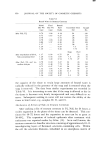
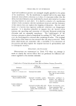
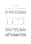
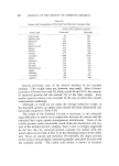
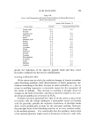
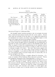
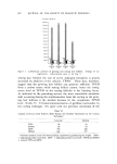
HUMAN HAIR FOLLICLES 909 tween the activities of telogen and anagen follicles. In the telogen stage, the enzyme activities in the upper half of the follicle are similar to those in the lower half, whereas in growing hair follicles the bulb generally has more enzyme activities than the external sheath. This change in activity is most dramatic in glucose-6-phosphate dehydrogenase (G6PDH), a key enzyme of the pentose cycle, where the impressive increases during ana- gen concur with the data obtained in the glucose-14C experiment, i.e., they both confirm the active participation of the pentose cycle in grow- ing hair follicles. As the hair follicles developed, phosphorylase activity increased con- comitantly in the external sheath and decreased in the bulb. Glycogen concentrations were elevated correspondingly, i.e., the anagen sheath contained 2.8 g glycogen/100 g dry weight, the bulb 0.44 (21). Thus, an inverse relationship exists between glycogen metabolism and biological activity. Glycogen accumulates in skin when the metabolism is sup- pressed (22, 23). How the mechanism of glycogen accumulation func- tions physiologically in the external sheath is still unknown, but the search for the mechanism should provide an interesting model for under- standing the metabolic control system in vivo. TESTOSTERONE METABOLISM In Vitro IN HUMAN HAIR FOLLICLES Wotiz et al. (24) have shown that human skin can metabolize testos- terone efficiently in vitro. Recently, Gomez and Hsia (25) incubated human skin with testosterone-4-nC and identified by paper chroma- tography and thin-layer chromatography such metabolites as andro- stenedione, 5a-dihydrotestosterone, 5a-androstanedione, androsterone, and epiandrosterone. The presence of an active catabolic pathway clearly suggests the physiological significance of skin, one of the largest organs in the human body. In order to study the etiologic factors for common baldness, we are concerned with the metabolic (catabolic) capacity of human hair follicles but not of whole skin. The possi- bility has recently been considered that sebaceous glands may also con- tribute to testosterone metabolism in hair follicles however, it will not be discussed here. Recent evidence suggests that 5a-dihydrotestosterone rather than testosterone is the active androgen in the target tissue. Bruchovsky and Wilson (26-28), for example, clearly demonstrated that in the prostate the predominant androgen is not testosterone but 5a-dihydro- testosterone and that 5a-dihydrotestosterone retained in the nuclear

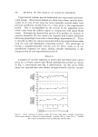
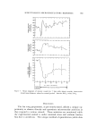
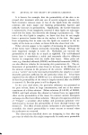
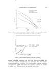
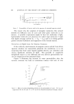
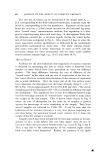
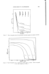
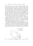
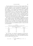
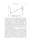
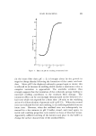
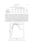
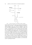
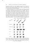
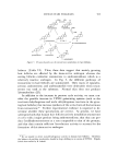
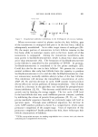
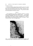
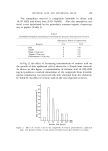
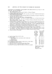
910 JOURNAL OF THE SOCIETY OF COSMETIC CHEMISTS fraction is not catabolized while that in the cytoplasmic fraction is rapidly broken down. Clinical evidence has shown that women with the testicular feminization syndrome are unresponsive to testosterone administration in spite of high testosterone levels corresponding to those of normal men (29). Since no 5oz-dihydrotestosterone is present in plasma, it must be formed from testosterone only in the peripheral tar- get tissue by a 5oz-reductase. In fact, a number of data on the skin of these patients indicate the lack or diminution of 5oz-reductase activity (27,30). The following experiments were designed to demonstrate the po- tential capacity of the hair follicles to convert testosterone to dihydro- testosterone. The problem was approached by in vitro experiment and measurement of 5a-reductase activity. Freshly plucked hair fol- licles were incubated for 30 to 60 rain at 37øC in a reagent mixture con- sisting of 0.02 •c and 0.1 vg of testosterone-•4C in a total volume of 5 •1 of Krebs bicarbonate Ringer solution containing 200 mg glucose/100 mi. In this in vitro experiment, no colactor was added to the incuba- tion medium because of the use of a fresh hair follicle that presumably generates TPNH from the added carbon source. For the 5a-reductase assays, the hair follicles were first freeze-dried to destroy the cell mem- brane and structure, and then the samples (15 to 300 vg) were incu- bated for 15 to 30 minutes at 37øC in 5 v1 of reagent mixture consist- ing of 0.02 vc and 0.1 vg of testosterone-•4C, 2mM TPNH, 2mM diphosphopyridine nucleotide (DPN), and 20mM tris-HC1 buffer pH 7.4. The latter in vitro experiment was designed to assay the optimal capacity of 5mreductase at physiological pH as well as the potential capacity of testosterone conversion to androstenedione. In the latter in vitro experiments, addition of cofactors is mandatory 5a-dihydro- testosterone was not formed without the addition of TPNH and andro- stenedione without added DPN (or TPN). In both types of experi- ments, testosterone and its metabolites were extracted with methanol/ chloroform (1/2: v/v) and isolated according to the method described by Gomez and Hsia (25) with slight modifications.* A typical resttit of the in vitro experiment is summarized in Fig. 3. In both growing and resting hair follicles, the major catabolic product is clearly androstenedione. The growing hair follicles metabolize testosterone faster than the resting hair follicles do (when they are * To be reported elsewhere in detail by S. Takayasu and K. Adachi.
Purchased for the exclusive use of nofirst nolast (unknown)
From: SCC Media Library & Resource Center (library.scconline.org)