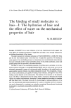
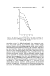
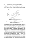
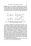
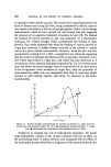
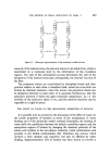
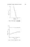
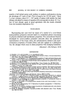
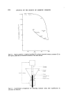
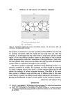
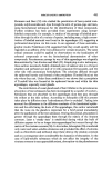
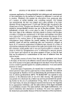
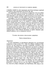
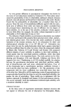
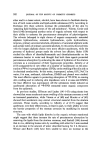
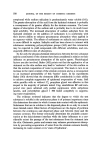
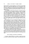
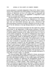
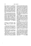
EFFECT OF SALT SOLUTIONS ON STRATUM CORNEUM 477 exists for the salt ions, then passage of sucrose through the lipid barrier should be facilitated for native stratum corneum if size was not the pre- dominant factor. It is suggested, therefore, that the ability of the corneum to retain its natural hygroscopic materials, when the lipid barrier is intact, is due, in some extent, to the size of the molecules which comprise the NMF. For reasons which have been discussed in the previous study (2) it has been suggested that the mechanical integrity of the corneum might reside in its intercellular junctions (junction = two adjacent cell membranes + desmosomes which act as intermediate adhesive material) rather than with- in the keratin filaments as had been assumed in the past (2). The present results, together with data obtained by Middleton (17) on the osmotic characteristics of the stratum corneum help to enhance this point of view. Middleton has shown that osmotic dehydration of stratum corneum occurs when it is bathed in a solution which has a higher osmotic pressure than the corneum cell contents. This will certainly be the case for the saturated solutions used in this study. Such a property can only be accounted for if significant amounts of the solutes do not penetrate into the corneum cells. If this is the case then the decrease in modulus values of the native corneum produced by the salt solutions in the present study must result from the action of these salts on the intercellular system furthermore, it is not too much of an extrapolation to suggest that the NMF is also located primarily in this region. It now only remains to consider the role of the lipids which allow pene- tration of small ions into the intercellular material but prevent the entry or removal of larger similar species. To this end, the salient features of a model for the junction between corneum cells are proposed in Fig. 4. The model consists of a protein/polysaccharide-rich central region (11) con- taining hygroscopic materials and isolated pools of lipid enclosed by the two adjacent cell membranes (lipid bilayers in association with proteins). The lipid bilayers of the cell membranes offer high resistance to the passage of charged species and water [complete hydration of the corneum requires •5 days (18)] and are considered to be responsible for the semi-permeable nature of the corneum. The large naturally-occurring hygroscopic molecules are retained within the central region of the junction by the lipid pools in the structure. That is to say, their removal by aqueous extraction is pre- vented due to steric factors. Extraction with lipid solvents removes all of the lipid and the hygroscopic substances are then readily removed with water, being leached out through the channels created by removal of the pools of lipid. The involvement of pools of lipid within the proteinaceous region of

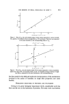
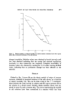
478 JOURNAL OF THE SOCIETY OF COSMETIC CHEMISTS cII CI2 O= Protein/polysaccharide matrix • =Lipid molecule Figure 4. Schematic diagram of corneum intercellular junction. CI, cell interior CM, cell membrane H, hygroscopic molecule. the junction is necessary to account for failure of the NMF to be removed by aqueous extraction while the small salt ions used here can enter the structure. It is envisaged that the initial entry of small ions into the central region of the intercellular junction occurs at the surface of the corneum where desquamation results in a breakdown of the lipid bilayer. After entry has been gained, these small ions can diffuse through the entire intercellular system which is continuous throughout the corneum. The above model is somewhat speculative and additional work is re- quired to establish its validity. For example, no effect should be observed on native corneum due to ions above a critical size but these should still reduce the modulus of solvent extracted corneum. The effect of the same ionic species at different water activities and of different salts at the same water activity (specific ion effect) should afford additional information re- garding the mechanism of action of such species on the corneum structure. (Received: loth March 1972) REFERENCES (1) Park A. C. and Baddiel, C. B. Rheology of stratum corneum I. A molecular interpretation of the stress-strain curve. J. Soc. Cosmet. Chem. 23 26 (1972). (2) Park, A. C. and Baddid, C. B. Rheology of stratum corneum II. A physico-chemical investigation of factors influencing the water content of the corneum. J. Soc. Cosmet. Chem. 23 36 (1972).
Purchased for the exclusive use of nofirst nolast (unknown)
From: SCC Media Library & Resource Center (library.scconline.org)