492 JOURNAL OF THE SOCIETY OF COSMETIC CHEMISTS between vasoconstrictor ability and clinical efficiency (82-84). The produc- tion of an area of anaesthesia by a topically applied substance could be used as a means of detecting percutaneous absorption, but it is even more subject to error than the vasoconstriction test (85). Other pharmacological parameters have been found useful in determin- ing percutaneous absorption and are still used occasionally in order to relate pharmacological action with rates of absorption measured by other tests. For example, changes in serum cholinesterase have been used to compare the toxicity of parathion and paraoxon after dermal application (54, 86, 87). Other criteria which have been used occasionally are death of test animals (so-called cutaneous LD50) from topically applied compounds or organ damage assessed histologically. Such an approach does not give an accurate measurement of percutaneous absorption but may be useful in order to obtain data on the dose levels at which certain compounds may produce systemic toxicity when applied topically. This approach was used by Wahlberg (74) in a comparative study of the systemic toxicity of mercuric chloride, cobaltous chloride and sodium chromate and is extensively used in determining the dermal toxicity of pesticides (88). It is not always possible to compare the results of percutaneous absorp- tion using an isotope-labelled compound with those obtained using other methods of measurement. In many instances the sensitivity of the analytical techniques employed is very much less than the radio-isotope techniques so that meaningful comparisons are difficult. Antibiotic assays using micro- biological techniques are sensitive and accurate and Vickers (89) compared the percutaneous absorption of sodium fusidate and fusidic acid, using such techniques with the result of absorption obtained by standard •4C-labelling techniques. Both by in vivo and in vitro methods, the results were found to be very close confirming the reliability of the radio-isotope techniques. The measurement or demonstration of skin absorption using biological effects is limited to compounds having a high biological activity. The use of this technique for cosmetic ingredients is therefore limited but is im- portant in the case of biologically active materials used in some permanent waving solutions or that will control bacteria on the skin, influence meta- bolism in such ways as to improve the texture or appearance of the skin, retard perspiration, or control dandruff. Autoradiography Autoradiography has been employed in the study of percutaneous

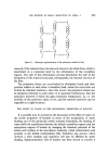
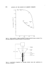
PERCUTANEOUS ABSORPTION 493 absorption in vivo (54, 91). This technique is useful in determining the presence of the compound in the various anatomical layers of the skin and in gaining some idea of the relative concentration. However, it is of limited value for quantitative investigations. Enhancement of penetration It is sometimes necessary to simulate 'conditions of use' where the skin surface may be exposed to injury and loss of its protective keratin expected. One method frequently employed is to remove keratin by the successive application and stripping of adhesive cellulose tape to the same cutaneous site. The number of consecutive strippings is usually about 25 (53, 92). The depth of epidermis removed by the adhesive is not uniform. Histological studies of tape removed after firm application revealed that in some areas the complete epidermal barrier is removed but in other areas only the merest trace of keratin (93). Other authors claim that a uniform separation of the keratin barrier is achieved by this method (29) so that the barrier is uniformly weakened. The skin barrier can be removed also by other methods. Recently Parekh et al (57) scarified the skin with a 'dull' razor to the point of localized bleeding in order to assess the absorption of sodium pyridinethione from damaged skin. Percutaneous absorption 'in vitro' methods In vitro measurements are found particularly useful in comparing the rates of diffusion of different compounds (74, 94, 95) and in obtaining some idea of the rate of the transepidermal passage of highly toxic substance prior to in vivo tests (42). They also provide a means of obtaining a better understanding of the factors that influence percutaneous absorption in vivo (9, 96) Both human and animal skin have been used for in vitro studies. Human skin is obtained at necropsy generally from the abdomen (42, 97) and from the abdomen, flanks or back in the case of animals (98, 99). Although the methods employed for the in vitro measurement of percutaneous absorption vary considerably in details, they follow a general pattern. The specimen of skin is trimmed to a suitable size and is mounted in a hollow chamber so that it divides the chamber into two compartments. The two surfaces of the skin are bathed in a suitable fluid. One of the fluids contains the test sub- stance. Passage through the skin is then measured either by the 'dis- appearance' of the test substance from one chamber, by its appearance in the other or by both methods combined. Full details of the construction of
Purchased for the exclusive use of nofirst nolast (unknown)
From: SCC Media Library & Resource Center (library.scconline.org)