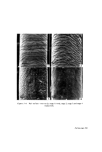
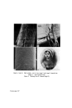
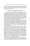
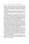
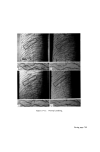
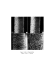
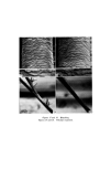
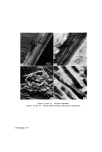
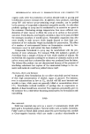
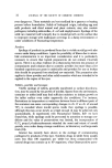
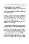
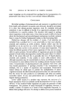
CHANGES IN SURFACE OF HAIR DUE TO COSMETIC TREATMENT 701 Finally, one of the clearest demonstrations of our present techniques has been in the examination of the split ends of naturally weathered human hair after the application of a polymer. The pairs of micrographs of Figs. 19-22 show that the disorganized cortical fibrils of the split ends are neatly replaced in the body of the hair fibre, restoring it to its original gross configuration. EXAMINATION OF THE FRAGMENTS BREAKING OFF HAIR AFTER COMBING AND MECHANICAL AGITATION The previous studies of hair surfaces have demonstrated that the scale edges of human hair break off with mechanical atrophy and it was therefore pertinent to examine the nature of the fragments released from the hair. The root end of a switch of untreated hair was thoroughly washed in distilled water with many rinses. After drying, one part of the switch was cut into pieces of 2 cm length, again washed thorougly with distilled water and then thoroughly shaken with distilled water in a flask on a laboratory shaker. Another part of the switch was combed (dry) with 2 000 strokes of a nylon comb and then rinsed in a small volume of water. The remainder of the switch was combed wet with 200 strokes of a nylon comb and this too was rinsed in a small volume of water. In each case the water was turbid and was therefore centrifuged. The various sediments were prepared for examination in the scanning electron microscope and also some embedded in epoxy resin, sectioned and stained for examination by transmission electron microscopy. Under the scanning electron microscope all the residues were seen to be composed of tiny platelets (Fig. 23) up to 5 gm in diameter and 0.3 gm thick. These are evidently fragments of the hair cuticle. In section (Fig. 24) the pieces are nearly all of one scale thickness and from their structure it would appear that they have been released mainly by cleavage along the cuticle cell membranes. A few of the pieces had also been released by cleavage through the endocuticle, a cystine-poor layer which constitutes one of the two major lamina which make up each cuticle cell (4). DISCUSSION Our experiments have shown that with treatments such as bleaching and perming and even with combing, the scale margins of human hair slowly chip away. The changes are relatively minor but nevertheless even with combing we can expect, and in fact do see, the gradual loss of the hair cuticle with time (i.e. from root to tip).

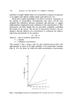
702 JOURNAL OF THE SOCIETY OF COSMETIC CHEMISTS For combing alone the rate of loss of cuticle was about 0.5 •tm after 2 000 comb strokes. Since the average length of each cuticle scale along the fibre axis is 40 •tm, some 160 000 comb strokes would be required to remove all the cuticle. The average rate of growth of human hair is 0.35 mm day -•, therefore the removal of all the cuticle at a distance of 40 cm from the scalp would require the subject to comb her hair at the rate of 150 comb strokes day 4. Clearly this is unlikely to happen, therefore the total lack of cuticle we have observed 40 cm from the scalp cannot have been due solely to combing, and other factors such as handling, exposure to sunlight, washing etc. must also contribute. That only small fragments chip from the edges of the scales occurs with such treatments as combing rather than the gross stripping away of the cells perhaps indicates that the cuticle is inherently brittle. This is not surprising in view of the fact that one of the major lamina of which each scale is composed (the exocuticle) is very highly cross-linked (containing some 30-40• cystinc w/w) (4). Since we have shown that cleavage occurs mainly along the cuticle cell membranes, and to a lesser extent in the sulphur-poor endocuticular lamina, it would appear that these components are mechanically the weakest in the cuticle. Indeed this confirms earlier observations (5) in which we have shown that bleaching and perming tend to extract small amounts of the cuticle cell membrane. ACKNOWLEDGMENTS We are indebted to Mr T. C. Hughes and Miss S. J. Williams and Miss P. McCarthy who have contributed to some of the work recorded here. Grateful thanks are also due to the trustees of the Jane Austen Society and particularly to Sir Hugh Smiley, Bt. and Miss E. Jenkins for permitting us to examine the lock of Jane Austen's hair and to reproduce Fig. 8. (Received: 13th April 1972) REFERENCES (1) Swift, J.A. New developments in electron microscopy. J. $oc. Cosmet. Chem. 22 477 (1971). (2) Austen, C. 'My Aunt Jane Austen'. 1867 manuscript republished by The Jane Austen Society, Chawton, Hants (1952). (3) Austen-Leigh, J. E. 'A Memoir of Jane Austen'. 1871, Richard Bentley & Sons, London. (4) Swift, J. A. The electron histochemistry of cystinc-containing protein in thin transverse sections of human hair. J. Roy. Microsc. Soc. 88 449 (1967). (5) Swift, J.A. Electron microscope studies of diffusion in human hair. Proc. 3rd Int. Cong. Wool Text. Res. Paris I 265 (1965).
Purchased for the exclusive use of nofirst nolast (unknown)
From: SCC Media Library & Resource Center (library.scconline.org)