748 JOURNAL OF THE SOCIETY OF COSMETIC CHEMISTS Alternatively, vital dyes may diffuse through the walls of the blood vessels in normal tissue, but fail to colour the tissue in areas of damage where the blood vessels have been destroyed as a result of severe burns (73). Early attempts at using dyes to detect the extent of cutaneous injury due to burns, involved the use of' a modified van Geisen's stain (74) or fluorescein (75). In human studies, vital dyes such as kiton green (76) and disulphine blue (73) injected intravenously, have been found useful in delimiting areas of trauma resulting from severe injury. Tempest (73) considered that the degree of extravasation of the dye was indicative of the severity of. the injury. Although these vital dye methods may be of. value in examining the extent of injury in patients wiith severe burns or crushing, they would appear to have limited application in detecting epidermal damage due to topical applications of. cosmetics, since a subject would be coloured blue or green by this treatment. This is undesirable. Dyes have been employed in animal experiments for detecting the extent of damage due to irritants. Apart from kiton green and disulphine blue as used in clinical studies, pontamine sky blue (77-79), Evan's blue (80) and trypan blue (47, 81) also have been used. In an attempt to quantify the extravasated dye as an index of the tissue damage, Lockett and Jarman (82) compared the intensity of the dye in the skin with a series of comparator cards, Miles and his colleagues (83) measured the diameter of' the zone of increased dye extravasation and expressed this in terms of the 'mean lesion diameter', whereas others made simple visual assessments of the dye intensity in the skin (47, 71, 84). Judah and Willoughby (81), however, believed that none of these methods was accurate enough to quantify an injury and they made extractions of the extravasated dye from tissue samples using standard analytical procedures (78, 80). This is not normally practicable in human studies but when biopsies are taken, these are more profitably examined histologically. The value of' dye extravasation techniques to assess injury which is not evident to the naked eye is questionable since in those studies where dyes have been used, the damage to the skin has been con- siderable and should have been detected unaided (71, 73, 76). Where biopsies can be taken and histological studies conducted, minor degrees of' epidermal damage can be assessed more accurately. Epidermal stripping studies As a means of detecting minor changes in the skin which escape recogni- tion with the naked eye, epidermal stripping may be of value. Anatomical

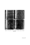
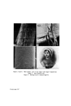
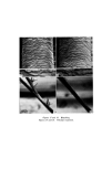
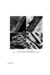
APPRAISAL OF METHODS FOR DETECTING PRIMARY SKIN IRRITANTS 749 studies using this technique date back to the seventeenth century when Marcello Malpighi and others, used warm water to separate the epidermis from underlying tissues in skin samples obtained at autopsy or by biopsy. More recent workers have used ammonia fumes (85) or enzymes (86) to obtain samples of epidermis. However, it is thought that the separation of the epidermis by these methods is likely to result in sufficient damage to obscure the effects due to treatment with an irritant. Another method for separating the epidermis from the dermis relies on the difference in the extensibility of the tissue. By stretching skin samples, the stratum corneum, epidermal cell layer and dermis can be separated (87-89). Although it is unlikely that any useful morphological information could be obtained from tissues prepared in this way, the technique may be suitable for biochemical studies where enzyme assays and analytical studies are contemplated. A technique which has been used for obtaining samples of epidermis from patients, employs blister formation. Blisters induced in the epidermo- dermal region of the skin using chemicals or suction technique, allow an excision of small samples of epidermis to be taken with a minimum of discomfort. 0.25/0 Cantharidin in acetone is capable of raising blisters on the skin of most parts of the body, but on the palms or soles of the feet, 0.55/0 is normally required (90). The extremely toxic nature of this chemical (91, 92) would preclude the use of this technique in routine dermatological studies of cosmetic materials. A method for the induction of blisters using a suction technique was described by Kustala and Mustakallio (93). They used an Angiosterrometer which was applied to the skin for about 3 h, during which time vesiculation and bulla formation occurred in the epidermo-dermal region. Having excised the required sample of epidermis, the remaining tissue of the blister may be pressed back on to its base, re-attachment being complete within 24 h. In this way a minimum of distortion and tissue damage is caused to the excised epidermis and suitable samples are obtained for histological or biochemical studies (94). A drawback with the method con- cerns the inability to induce epidermo-dermal blisters in those skins exhibit- ing pronounced acantholytic changes. In this instance the blisters form in the intra-epidermal region. This observation suggests that the technique is limited in its application to obtaining epidermal samples from skin which is only mildly damaged or is normal. However, the method has been found useful recently in the study of wound healing (95). Adhesive tape has been used for several years to remove the keratinized
Purchased for the exclusive use of nofirst nolast (unknown)
From: SCC Media Library & Resource Center (library.scconline.org)