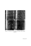
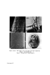
750 JOURNAL OF THE SOCIETY OF COSMETIC CHEMISTS cells from human and animal skins for studying epidermal regeneration (38, 96) and the role of the epidermis in percutaneous absorption (97). Only on a few occasions have workers examined the cells removed for comparative anatomical purposes, or for detecting changes due to the effects of topically applied substances (98). Since Wolf (99, 100) originally described this technique, the method has been modified on several occasions (98, 101, 102). The cells on the surface of the skin are removed using a suitable adhesive tape which is applied using rollers to ensure that complete ad- hesion is achieved. The tape is rapidly removed and with it are the epidermal cells, which can then be stained and examined microscopically. Repeated applications of tape to the skin allow successive layers of cells to be removed down to the Malpighian layer. Although no reports seem to be available to show the value of this technique in examining skins treated topically with irritants, the method is potentially useful for giving information about changes such as hyperkeratosis or parakeratosis, which are frequently present in this type of investigation. Features which make the technique attractive are that it is simple in design and requires little specialized apparatus. The chief disadvantages of the tape stripping technique concern the hairiness of the skin and the irregularity of the layers of the epidermis. With hairy skin, clipping is important and was reported to be preferable to wax depilation or shaving, both of which are liable to result in damage to the superficial layers of the epidermis. The unevenness of the skin, due to the presence of dermatolglyphics and epidermo-dermal ridging, might lead to difficulty in interpretation, since single strippings are likely to contain cells from several layers. This difficulty may be minimized by taking strippings only from small areas of skin. Using a tape stripping technique, it is possible that minor epidermal changes which escape recognition with the naked eye will be detected. It is suitable for use in connection with the patch test, when it is not convenient to take skin biopsies for histological examination. Enzyme studies Histochemical and biochemical studies of enzyme systems have been found useful to detect changes in epidermal metabolism resulting from treatment with hydrocarbons and other agents applied topically. Biochemic- ally, hydroxysteroid dehydrogenase (103), arginase, i.e. L-arginine-amidino- hydrolase (104-107), and protocollagen proline hydroxylase (108), have

APPRAISAL OF METHODS FOR DETECTING PRIMARY SKIN IRRITANTS 751 been studied in biopsied skin samples. Alteration in the levels of the enzymes were related to changes in the steroid metabolism of the skin, the degree of keratinization and the dermal collagen synthesis respectively. An important disadvantage with the techniques used is that sufficient skin has to be biopsied to enable several biochemical assays to be performed using different substrates. A limitation of the hydroxysteroid dehydrogenase assay is that only those steroids with hydroxyl groups may be determined and if specific assessments are required only steroids with a single hydroxyl group can be used. The assessment makes no allowance for alterations in cutaneous steroid metabolism resulting from endocrine disturbances or from alterations in local concentrations due to the action of the circulatory system. The assay for arginase, using the method devised by Rossmiller and Hoekstra (109) and the assay for protocollagen proline hydroxylase also would appear, on available evidence, to be of limited application and to be an unreliable guide to irritation. Another enzyme which has been investigated as a possible guide to the irritancy of cosmetic materials, is saccharase (110). The test was based on the inhibition of the enzyme by some anionic detergents which were known to irritate the skin by damaging the keratin. It would seem, however, that the same physical properties of those detergents which are responsible for damaging the keratin, would also denature other proteins including the enzyme saccharase. The choice of an enzyme which is not normally present in the skin, would seem to make this test for cutaneous irritants of rather questionable validity. Whereas biochemical assays of enzyme levels in tissue samples seem to be an unreliable guide to tissue damage not visible with the naked eye, the histochemical demonstration of enzymes is likely to be of more value. Although few studies of this type have yet been reported, Reid and Jarrett (111) have shown that vitamin A treatment results in an increase in the level of lysosomal hydrolases in the stratum granulosum. Similar changes were noted in skin treated with the irritant-cetyl trimethylammonium bromide (112). In these studies, changes in enzyme levels were present before changes were identified histologically. Thus changes in enzyme levels appear to be more sensitive indicators of tissue damage than histological techniques and would appear to offer a considerable advantage over the in vitro assays referred to above. Keratin denaturatiot• When a substance irritates the skin, it may damage the epidermal keratin
Purchased for the exclusive use of nofirst nolast (unknown)
From: SCC Media Library & Resource Center (library.scconline.org)