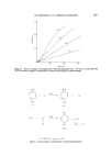
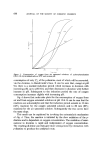
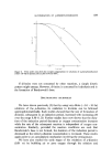
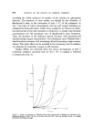
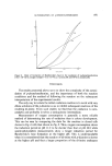
752 JOURNAL OF THE SOCIETY OF COSMETIC CHEMISTS of the stratum corneum and the stratum lucidum, or it may penetrate deeper into the epidermis, thereby bringing about cytotoxic changes (113). With the exception of a thin layer of surface debris and lipids secreted by the sebaceous glands, the keratin layer constitutes the outward line of the defence to the skin to potentially harmful substances. It is possible that information as to how a substance alters the keratin layer and the essential substances con- tained in it, may be of considerable value in detecting damage which is not visible to the naked eye. The horny layer of mammalian skin is composed mainly of the protein keratin, water and hygroscopic substances such as lipids, mucopolysacchar- ides, amino acids and sugars (114-117). Blank (114) maintained that for human skin to retain its supple nature it should contain at least 10}/o water. Although the removal of the hygroscopic substances apparently does not affect the ability of the skin to be rehydrated, the water is readily lost at low humidities and high temperature (118). An increase in the number of free sulphydryl groups in the keratin is evidence of denaturation resulting from a breakage of the disulphide link- ages in the amino acid cystinc (119, 120). Colorimetric methods have been used to determine these groups (119, 121) and the values used as an index of keratin denaturation (121, 122). Irritants such as detergents may free and elute essential amino acids or lipids from the keratin layer (123). To investigate such an effect, an apparatus consisting of a double-walled chamber is used. The suspected irritant dis- solved in a suitable solvent is placed in the chamber in contact with the skin stimulated with electrically operated teflon rollers for a test period of 15 min. At the end of the treatment, the fluid plus the amino acids and other substances eluted from the skin is analysed using standard techniques. Although the original technique was designed to examine the effect of detergents, it could be readily adapted to examine the effects of other irritants in a suitable solution. The test period could be varied according to the potency of the test substance. The removal of lipids from the epidermis has been studied using open type techniques (124). In one such study, animals were anaesthetized and dipped up to their necks in a test solution such as n-hexane and the removed lipids identified using thin layer chromatography. In a human study, the effects of some organic solvents on the scalp were examined using a simple washing procedure. The eluted lipids were characterized and quantified biochemically (125, 126). The design of these techniques appears to be relatively simple and enables them to be readily available for use hz vivo

APPRAISAL OF METHODS FOR DETECTING PRIMARY SKIN IRRITANTS 753 with animals or humans. Suitable reference standards are essential in order to relate new substances to others of known irritancy. Although there appears to be some controversy as to the main 'barrier' to the penetration of substances into the skin, there seems to be a general agreement that the stratum corneum is largely responsible (12, 97, 113, 127). Thus, any agent that damages the stratum corneum is liable also to impair the barrier function of the skin and make the skin more permeable to foreign substances. Tests designed to show how an agent alters the permeability of the skin will on this reasoning also demonstrate how the substance alters the keratin layer. A detailed review of methods designed to show how topically applied substances alter the permeability of the skin, by impairing the barrier function of the keratin layer, appears elsewhere (128). Discussion In an evaluation of the safety of substances likely to come into contact with the human skin, the patch test, firstly using animals and subsequently with human volunteers, on available evidence seems to provide useful information as to whether a substance is irritant or not. However, as Idson (25) emphasized, no single test is entirely satisfactory and more sensitive indicators for tissue damage are required. The main drawbacks of the patch test concern the occluded conditions required for testing and the subjective assessment used for evaluating the cutaneous reactions. These disadvan- tages are as applicable in animal tests as they are in clinical trials (11, 54). Whereas in animal tests, discrepancies due to the method of scoring can be overcome using histological techniques, such a procedure is not normally practicable in clinical trials. It would seem important that when human trials are conducted, they should be carried out under strict medical supervision (59). The objection to using the patch test only under occluded conditions is remedied by treating some subjects with open patches where the test compound is applied to the uncovered skin. An important source of error, illustrated by Kligman (11), is that certain substances may produce damage which is not evident at an early stage but is detected only when it has attained an advanced stage and necrosis is present. On this evidence, simple visual assessments of areas of treated skin are not entirely satisfactory and more sensitive markers for tissue damage are required. The use of vital dyes is not thought to be satisfactory for delimiting areas of damage which are not already visible to the naked eye. However, a
Purchased for the exclusive use of nofirst nolast (unknown)
From: SCC Media Library & Resource Center (library.scconline.org)