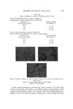
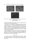
J. Soc. Cosmet. Chem., 24, 609-622 (September 16, 1973) Innovative Scanning Electron Microscopic Techniques for Evaluating Hair Care Products SAL P. DxBIANCA, M.A.* Synopsis-The utilization of microscopy in studying human hair is briefly reviewed. Reasons for selecting the SCANNING ELECTRON MICROSCOPE (SEM) over the transmission electron microscope and the optical microscope are discussed. The use of the SEM in evaluating HAIR CARE PRODUCTS is then described. A new technique employing a ROTATING HAIR STAGE, specially designed and fabricated for this study, is presented. The procedure devised allows one to view hair in the SEM while still attached to the panelist's head. The technique is nondestructible to the hair, per- mitting the study of sequential treatments on the same hair. For example, the evaluation of a shampoo on the hair after 0, 5, 10, and 20 treatments is now possible. The hair is removable from the SEM as many times as required for treatment without the necessity of cutting the hair from the scalp. In addition, the apparatus allows for complete axial rotation of the hair in the SEM. The functionality of two hair care products (a shampoo and a conditioner) is demon- strated using this technique. MICROGRAPHS of hair damages before and after treatment are categorized and numerically rated. The difference ratio was devised as an index to measure the degree of improvement of damaged sites. INTRODUCTION In the past, evaluation of hair care products centered around subjective beauty salon studies. Recently, however, the value of the scanning electron microscope (SEM) in revealing the effects of hair preparations has come to the forefront. This report summarizes part of our investigations into this area and reveals how to employ the scanning electron microscope as a tool to dem- onstrate the functionality of a hair product. Before giving the details of this *The Mennen Co., Morristown, N.J. 07960. 6O9

610 JOURNAL OF THE SOCIETY OF COSMETIC CHEMISTS investigation it is pertinent to briefly describe the capabilities and limitations of the three microscopic techniques available and demonstrate why the SEM was chosen. MICROSCOPY OF HUMAN HAIR The scanning electron microscope has overcome many limitations of optical microscopy and conventional transmission electron microscopy in elucidating the structure of human hair. Using the optical microscope, one views a pattern of light and dark areas produced by the reflection or passage through a thin slice of the specimen. Al- though hair can be viewed without interferences, while maintaining its natu- ral colors, only those parts that lie in the same plane can be reproduced sharply. Also, since light in the visible range of the spectrum is the energy source, the diameter of each part reproduced must be larger than the wave- length of light. Only at low magnifications (below 200 diameters) is the light microscope useful for showing the shape and depth of hair. In order to avoid this problem, early studies of hair involved the viewing of very thin hair disks. Only limited information can be drawn from this tech- nique, for only an extremely thin section of the specimen can be viewed. The conventional transmission electron microscope (TEM) far exceeds the magnification of the optical microscope. This enormous magnification (up to 200,000x ) allows the study of details which never appeared in the optical microscope. The TEM image is produced by monochromatic electrons that have illuminated a specimen which transmits or scatters the electrons. Once past the specimen, the electrons are then focused on a screen or sheet of film magnifying the image. What one sees is a two-dimensional pattern of light and dark areas produced by passage of electrons through the ultrathin speci- men. The use of ultrathin specimens results in an extremely low definition of depth. Since the transmission electron microscope no longer works with light but electrons, the images produced are not colored. Sample preparation for the TEM is complex and time consuming. Most bio- logical salnples are replicated. Acetyl cellulose or a similar material is placed over the specimen which is wetted with methyl acetate. After solvent volatili- zation the replica is carefully peeled off. This replica, or a second replica, is now shadowed. In this process a heavy metal such as platinum or gold is evaporated in vacuum on the sample surface. What we see then is not the sample but a two-dimensional silhouette of the metal deposits on the hair. Eliminating the optical microscopy problem of narrow depth of field and the transmission electron microscope limitations of extensive specimen preparations, the SEM has gained preference in today's research endeavors. It is an extremely versatile instrument revealing the exact topographical struc- ture of the specimen. In normal operation, the magnification range extends from 30x to 200,000x. The high depth of focus, a bonus characteristic of
Purchased for the exclusive use of nofirst nolast (unknown)
From: SCC Media Library & Resource Center (library.scconline.org)