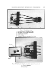
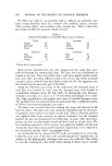
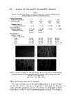
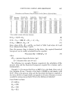
616 JOURNAL OF THE SOCIETY OF COSMETIC CHEMISTS No effort was made to use panelists •vith or without any particular hair types (virgin, bleached, dyed, etc. ), textures (fine, medium, coarse), amounts (thin, average, thick), and condition (oily, normal, dry). Table I shows the percentages of different categories finally selected. Table I Statistical Breakdown of Panelists' Hair Used in Testin ga Types Amounts Virgin 67 Thin 12 Bleached 25 Thick 41 Dyed 8 Average 47 Textures Condition Fine 46 Oily 47 Medium 50 Dry 2 Coarse 4 Normal 51 "Figures given as percentages. Each of four selected hairs was color tagged near the scalp, then care- fully fed through the rotating hair stage. The hairs were not conditioned or treated in any way. They xvere held in place with silver-plated wooden tooth- pick ends. After providing suffleient slack in the device, the rubber grommet xvas put in place, with the four hairs fitting xvithin the slit. The apparatus was then bolted into the open side post of the SEM. Using the television scan mode of the instrument, the damaged areas of each hair xvere located. In some eases the damaged areas were located at considerable distances along the hair shaft. To photograph such damages, montages consisting of as many as 8-10 individual photographs were made. Also, high magnification micrographs were made of selected damaged areas. The panelist's hair was removed from the instrument after a suffleient number of "before" treatment micrographs xvere taken. The hairs xvere removed from the rotating hair stage and allowed to fall back into place. The hairs were now randomly distributed and for all practi- cal purposes were similar to all others on the head. That is, when the product was applied, these hairs received no special treatment. For the shampoo product, the hair was shampooed (txvo latherings) six times. After each sham- pooing, the hair was thoroughly rinsed and dried with an electric drier. The ha r conditioner product, a leave-on type, xvas applied to slightly wet hair and electrically dried. After treatment the panelist's tagged hairs were returned to the instrument and the "after" micrographs taken. By viexving the hair by means of the TV mode and rotating it, we were able to find the exact area of the hair shaft and photograph the repaired area. In addition to the photographs, videotape data were also collected. This ability to view the scanning of a hair shaft while rotating it allows one to visually

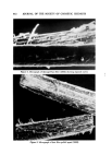
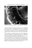
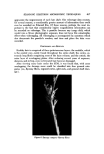
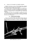
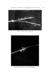
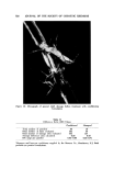
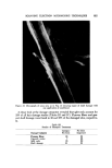
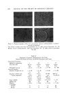
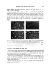
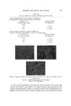
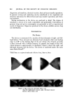
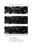
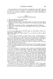
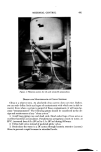
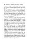
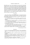
SCANNING ELECTRON MICROSCOPIC TECHNIQUES 617 appreciate the improvement of each hair shaft. The videotape data contain, for several reasons, a considerably greater amount of information than could ever be recorded on Polaroid film. Of these reasons, perhaps the most im- portant is the fact that considerably higher magnification information can be recorded on videotape. This is possible because any image drift, which would ruin a 50-sec photographic exposure, does not have this catastrophic effect when videotaping. All videotaping is accompanied by narration which also documents the panelist's number, and time and place the data were recorded. DISCUSSION AND I•ESULTS Healthy hair is composed of three proteinaceous layers: the medulla, which is the central core-rarely found throughout the entire shaft the cortex, ex- tremely long fibrils comprising most of the hairs volume and the cuticle, the outer layer of overlapping plates. After enduring several years of exposure, abrasion, and styling, even well-treated hair becomes damaged. After viewing many hairs under the SEM, it was found th.at, with some overlapping, the damage areas could be classified into four general cate- gories (i.e., flyaway fibers, exposed cortex, split ends, and general shaft dam- age). ..•.• ..•:• ..... ß .:•...1.•: .:..... "? .7!:': ß ,. } ß •. ß ..•. • :? :: Figure 6. Damage category-flyaway fibers
Purchased for the exclusive use of nofirst nolast (unknown)
From: SCC Media Library & Resource Center (library.scconline.org)