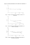
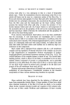
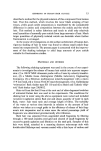
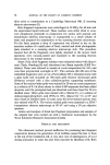
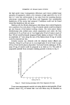
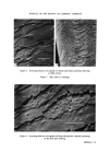
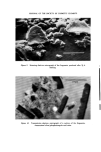
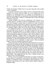
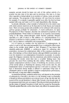
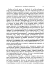
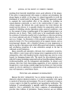
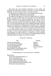
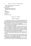
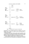
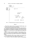
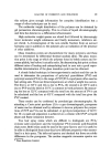
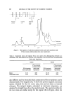
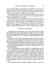
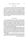
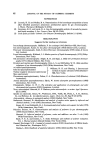
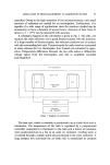
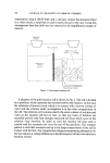
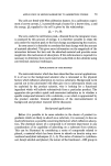
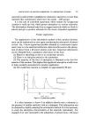
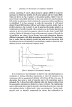
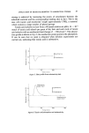
14 JOURNAL OF THE SOCIETY OF COSMETIC CHEMISTS overlay each other in a way analogous to that in a stack of disposable plastic cups. Under the transmission electron microscope some five to ten cuticle cell layers can be seen in a transverse section of the fibre (Fig. 1). From such electron micrographs it is clear that each cell is laminated, being divided into two principal components, the exocuticle (at the greater radius from the fibre axis) and the endocuticle (at the lesser radius). Similar sections but stained with silver salts (Fig. 2) show the presence of two further laminae within each cell: the A-layer which is a sub-component of the exocuticle and is at the periphery of the cell on the outer-facing aspect and the inner-layer which is between the endocuticle and the periphery of the cell on the inner-facing aspect. From electron histochemical observations (1) it has been established that the exocuticle, A- and inner-layers are exceedingly rich in cystine whereas there is little or no cystine in the endocuticle. In addition, since the endocuticle stains intensely with dodecatungstophosphoric acid (a trivalent anion of a heteropoly acid) and with uranyl acetate, we believe this indi- cates that acidic and basic amino acid residues are in relatively high con- centration in this component. Each cuticle cell is separated from adjacent cells by a cell membrane complex and the regularity of internal structure of the complex can be seen at high magnification in transmission electron micrographs of dodeca- tungstophosphoric acid-stained transverse sections of hair (Fig. 3) (2). The overall thickness of the complex lamina is about 28 nm. The central dark- staining portion, which is about 18 nm thick, is considered to be an inter- cellular cement composed of protein or polysaccharide, and is generally referred to as the delta-band. On either side of this is a narrow layer some 2.5 nm thick which cannot be stained and is thought to be the lipid com- ponent of the cell membrane from each cell (2). Electron microscope observations of human hair cuticle dearly reveal a multiplicity of sub-components. Considerable variation in the chemical composition of these various laminae may therefore be expected. ISOLATION OF CELLS Many methods have been described for the isolation of different cell types from mammalian keratin fibres. A large number of these have relied upon chemical treatments of the fibre (3-6) and are clearly to be avoided where further detailed chemical studies are required of the isolated cells. In the case of fibre cuticle Wolfram and Lindermann (7) have recently

JOURNAL OF THE SOCIETY OF COSMETIC CHEMISTS half s cortex Figure 1. Transverse section of human hair cuticle. Phosphotungstic acid stain. Figure 2. Transverse section of cuticle. Silver-methenemine stain. Figure 3. High magnification electron micrograph of sectioned cuticle cell membranes. Phosphotungstic acid stain. (Facing p. 14)
Purchased for the exclusive use of nofirst nolast (unknown)
From: SCC Media Library & Resource Center (library.scconline.org)