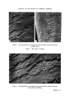
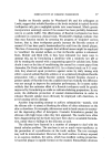
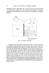
CHEMISTRY OF HUMAN HAIR CUTICLE 15 described a method for the physical isolation of this component from human hair. That this method, which involves the razor blade scraping of hair tresses, yields good cuticle preparations is manifested by the considerable differences in amino acid composition between the cuticle and the whole hair. This method is rather laborious and tends to yield only relatively small quantities of assuredly pure cuticle from large amounts of hair. Much larger quantities of physically isolated cuticle are desirable where further fractionation is envisaged. In the course of investigations on the surface architecture of human hair, vigorous shaking of hair in water was found to release small cuticle frag- ments into suspension (8). The present paper is concerned with the improve- ment of this shaking technique to yield large amounts of pure cuticle suitable for fractionation studies. MATERIALS AND METHODS The following shaking equipment was used in the course of our experi- ments to investigate the release of human hair cuticle into aqueous suspen- sion: (1) a 100 W MSE ultrasonic probe with a 9 mm tip velocity transfor- mer, (2) a Mickle tissue disintegrator (Mickle Laboratory Engineering Company), (3) a Silverson multipurpose mixer/homogenizer with a micro 15-9 mm head, (4) a Cenco-Virtis 45 macrohomogenizer, (5) a Gallenkamp 'wrist-action' flask shaker type SD 110 and (6) a Baird and Tatlock 'ellip- told' flask shaker type 330/0012. Hair cut from the first 10 cm at the root end of ether-degreased switches of untreated Italian hair was used in the experiments. The conditions for shaking hair in water using the above equipment were varied and included variation in agitation speed, flask size and shape, quantity of water in each flask, water: hair mass ratio and average length of fibres. The turbidity of the water at various time intervals in relation to the amount of hair shaken was taken as a rough guide to the efficiency of release of hair frag- ments into suspension and in some cases the actual percentage yield of fragments was determined gravimetrically after filtration. Bulk hair was separated from suspended small fragments by filtering through a 100 mesh stainless steel grid and cleared of small fragments by successive gentle agitation and filtration on the steel grid. Samples of this hair were dried, attached to a scanning electron microscope stub with double-sided Sellotape and vacuum coated with a thin layer of carbon and

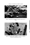
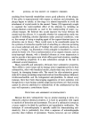
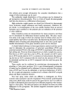
16 JOURNAL OF TIlE SOCIETY OF COSMETIC CHEMISTS silver prior to examination in a Cambridge Stereoscan Mk. II scanning electron microscope (8). Hair fragment suspensions were centrifuged at 38 000 g for 60 min and the supernatant liquid removed. These residues were either dried in vacuo over phosphorus pentoxide in preparation for amino acid analysis and transmission elec[36n microscopy or resuspended in a small volume of water and prepared for examination in the scanning electron microscope. For the latter purpose a drop of suspension was allowed to dry on the emulsion surface of a small piece of fixed, washed and dried photographic plate attached to a scanning electron microscope stub. This procedure ensured that all the fragments were lightly attached to the mount. Such specimens were vacuum-coated and examined in the scanning electron microscope in the normal manner. Some of the dried fragment residues were intimately mixed with Spurr's resin (Taab, Reading) (9) and introduced into Beam capsules (LKB Pro- dukter). These were allowed to remain at room temperature for 24 h and were then polymerized overnight at 65 ø. Thin sections (60-100 nm) of the embedded fragments were cut on a Porter-Blum MT-2 ultramicrotome with a glass knife and mounted on 100 mesh gold electron microscope grids (Polaron) covered with a thin collodion/carbon supporting membrane. Some of the section-laden grids were stained for 20 min at room temperature in a solution of 0.1 M silver nitrate to which 0.880 ammonia had been added dropwise until the precipitate had just dissolved and then rinsed for 30 s in distilled water. Other grids were stained with a filtered 25/0 w/v solution of dodecatungstophosphoric acid (PTA) in 505/0 ethanol for 3 h at 65 ø and then rinsed in distilled water. In some cases the silver-stained sections were also stained with PTA. The various stained grids were examined in a JEM 7 transmission electron microscope at 80 kV and using a 50 !xm objective aperture. Amino acid analyses of dried hair fragment preparation and samples of the original hair were carried out with a Technicon Autoanalyser by the Wool Industries Research Association (Leeds). RESULTS AND DISCUSSION The ultrasonic method proved ineffective for producing hair fragment suspensions because the generation of air bubbles caused the hair to float at the top of the irradiation cell. A very slow rate of disintegration of wool during sonication in water has also been reported by Bradbury (10). Both
Purchased for the exclusive use of nofirst nolast (unknown)
From: SCC Media Library & Resource Center (library.scconline.org)