Micro-organisms on human skin 615 15 Stringer, M. F. and Marpies, R. R. Ultrasonic methods for sampling human skin micro-organisms. Brit. J. Dermatol. 94 551 (1976). I6 Kligman, A.M., Leyden, J. J. and McGingley, K. J. Bacteriology. J. Invest. Dermatol. 67 160 (1976). 17 Smith, R. F. Comparative enumeration of lipophilic and nonlipophilic cutaneous diphteroids and cocci. Appl. Microbiol. 19 254 (1970). 18 Voss, J. G. Effects of an antibacterial soap on the ecology of aerobic bacterial flora of human skin. AppL Microbiol. 30 551 (1975). 19 Bibel, D. J. and Le Brun, J. R. Changes in cutaneous flora after wet occlusion. Can. J. Microbiol. 21 496 (1975). 20 Green, J. H. and Ronsivalli, L. J. A cylinder template method for sampling fish surfaces. J'./lppl. Bacteriol. 43 171 (1977). 21 Williamson, P. and Kligman, A.M. A new method for the quantitative investigation of cutaneous bacteria. J. Invest. Dermatol. 45 498 (1965). 22 Troller, J. A. Model system for the investigation of dandruff. J. Soc. Cosmet. Chem. 22 187 (1971). 23 Brewer, J. H. Safe self-contained carbon dioxide-hydrogen anaerobic system. •lppl. MicrobioL 44 985 (1966). 24 Prince, H. N. and Rodgers, J. A. Studies on the aerobic axillary microflora employing a standardized swabbing technique. Cosmet. Perfum. 89 25 (1974). 25 Montes, L. F. and Willborn, W. H. Location of bacterial skin flora. Brit. J. Dermatol. 81 Suppl. 1, 23 (1969). 26 Evans, C. A. Persistent individual differences in the bacterial flora of the skin of the forehead: numbers of propionic bacteria. J. Invest. Derrnatol. 64 42 (1975). 27 Noble, W. C. Dispersal of organisms from human skin. Cosmet. Toiletries 92 38 (1977). 28 Kloos, W. E. and Musselwhite, M. S. Distribution and persistence of staphylococcus and micro- coccus species and other aerobic bacteria on human skin. •lppl. MicrobioL 30 381 (1975). 29 Bibel, D. J. and Lovell, D. J. Skin flora maps: a tool in the study of cutaneous ecology. J. Invest. Dermatol. 67 265 (1976).

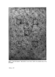
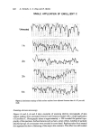
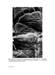
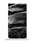
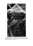
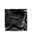
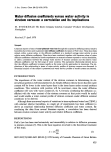
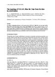
J. Soc. Cosmet. Chem. 29 617-624 (1978) Short term effects of emollients and a bath oil on the stratum cornoum* S. NICHOLLS, C. S. KING and R. MARKS Department of Medicine, Welsh National School of Medicine, Heath Park, Cardiff Received 24 August 1977 Synopsis Recently devised techniques were employed to detect and measure surface changes and structural altera- tions in the stratum comeurn after single applications of both oil in water emollients and a dispersion type of bath oil. Flattening of the surface contours of the skin and 'plumping' of individual corneocytes were apparent on replicas of the skin taken from treated areas and suggested hydration. These effects were most apparent when replicas were examined by scanning electron microscopy and by surface con- tour tracing. Small changes observed in skin surface blopsies from treated areas may reflect alterations in the internal structure of the top layers of stratum corneum. An increase in skin furrow width was also detected in photographs of the surfaces of treated skin. INTRODUCTION Considerable symptomatic relief for patients with itching and scaling dermatoses is obtained by measures designed to increase hydration of the stratum corneum (SC). Apart from symptomatic relief this type of therapy may actually hasten recovery by the reduction of fissuring, excoriation and scaling. In this study, we have attempted to assess changes in the surface structure of skin after single treatments with either water con- taining a dispersion type of bath oil, or one of two oil in water emulsion emollients using techniques developed by our department. MATERIALS AND METHODS Materials used Bath Oil A. Alpha Keri © (oil soluble fraction of lanolin, mineral oil and non-ionic emulsifiers), Westwood Pharmaceuticals. Emollient B. Keri lotion © (kerohydric © emollient, non-ionic emulsifiers), Westwood Pharmaceuticals. Emollient C. Oil of Ulay © (lipid particles in an aqueous phase), Garsalle Division of Richardson-Merrell, Ltd. The materials were supplied by the manufacturers and employed according to their instructions. Skin surface biopsies (SSBs) Samples of SC were taken onto glass microscope slides with a cyanoacrylate adhesive * Based on a paper read at the European Society for Dermatological Research, Amsterdam, May 1977 0037-9832/78/1000-0617 $02.00 ¸ 1978 Society of Cosmetic Chemists of Great Britain 617
Purchased for the exclusive use of nofirst nolast (unknown)
From: SCC Media Library & Resource Center (library.scconline.org)