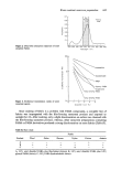
J. Soc. Cosmet. Chem. 29 617-624 (1978) Short term effects of emollients and a bath oil on the stratum cornoum* S. NICHOLLS, C. S. KING and R. MARKS Department of Medicine, Welsh National School of Medicine, Heath Park, Cardiff Received 24 August 1977 Synopsis Recently devised techniques were employed to detect and measure surface changes and structural altera- tions in the stratum comeurn after single applications of both oil in water emollients and a dispersion type of bath oil. Flattening of the surface contours of the skin and 'plumping' of individual corneocytes were apparent on replicas of the skin taken from treated areas and suggested hydration. These effects were most apparent when replicas were examined by scanning electron microscopy and by surface con- tour tracing. Small changes observed in skin surface blopsies from treated areas may reflect alterations in the internal structure of the top layers of stratum corneum. An increase in skin furrow width was also detected in photographs of the surfaces of treated skin. INTRODUCTION Considerable symptomatic relief for patients with itching and scaling dermatoses is obtained by measures designed to increase hydration of the stratum corneum (SC). Apart from symptomatic relief this type of therapy may actually hasten recovery by the reduction of fissuring, excoriation and scaling. In this study, we have attempted to assess changes in the surface structure of skin after single treatments with either water con- taining a dispersion type of bath oil, or one of two oil in water emulsion emollients using techniques developed by our department. MATERIALS AND METHODS Materials used Bath Oil A. Alpha Keri © (oil soluble fraction of lanolin, mineral oil and non-ionic emulsifiers), Westwood Pharmaceuticals. Emollient B. Keri lotion © (kerohydric © emollient, non-ionic emulsifiers), Westwood Pharmaceuticals. Emollient C. Oil of Ulay © (lipid particles in an aqueous phase), Garsalle Division of Richardson-Merrell, Ltd. The materials were supplied by the manufacturers and employed according to their instructions. Skin surface biopsies (SSBs) Samples of SC were taken onto glass microscope slides with a cyanoacrylate adhesive * Based on a paper read at the European Society for Dermatological Research, Amsterdam, May 1977 0037-9832/78/1000-0617 $02.00 ¸ 1978 Society of Cosmetic Chemists of Great Britain 617

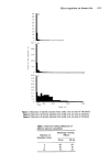
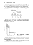
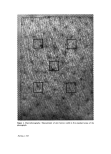
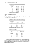
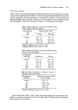
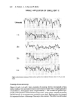
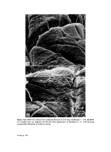
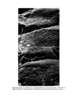
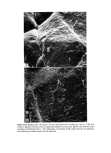
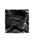
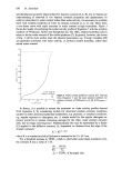
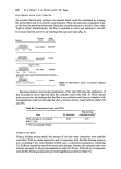
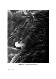
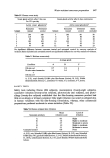
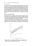
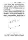
618 S. Nicholls, C. S. King and R. Marks (Permabond Staident Ltd) using the skin surface biopsy technique to reveal the ruptured internal surface of' the outer SC (1). Skin surface replicas Negative impressions of the skin surfaces were taken with standard Silflo impression material (J. & S. Davies, London). Positives of the skin surface were subsequently obtained from the 'negative' by covering them with DPX (R. A. Lamb, London) histological mounting medium (2). The negatives with DPX were then placed in a desiccator for 5 or 6 h or left overnight at room temperature before separating the 'positive' from the 'negative'. Scanning Electron Microscopy (SEM) Replicas and SSBs were mounted on stubs and coated with gold in a Polaron ES000, sputter coater, and examined in a Cambridge Stereoscan S2 scanning electron microscope at approximate magnifications of x 200 and x 1000. Macrophotography Photographs of the skin surface were taken using a 35 mm camera fitted with a 105 mm lens on a bellows attachment and an electric ring flash. Negatives were printed to give a final magnification of x 8. Skin furrow widths were measured with an eye piece graticule ( x 8) by taking measurements in each of five standard areas in the photographs (Fig. 1), and averaging the values for the five areas. Surface contour measurement of SSBs and replicas SSBs and replicas were examined in an apparatus (3) designed for measuring surface contours (Suffometer, Planer Products, Ltd). The surfometer employs a stylus to tra- verse a test surface, the contours of which are traced on moving chart paper. Skin surface biopsies (SSBs) The areas under the curves of the surfometer tracings were measured with a planimeter and the mean peak height calculated for a representative 10 cm length of trace. Skin surface replicas Only very thin DPX positives were suitable for surface contour tracing. When separated from the negatives they were left to harden at room temperature. The DPX positives were then fixed to glass slides and the surface contours measured in our surfometer. The area described by the surface contour in 10 cm length of trace was measured with a planimeter. (For the Oil of Ulay study (C) tracings of 8.5 cm length of trace were used and the total length of contour was also measured using a curvimeter). Ambient conditions Laboratory personnel acted as volunteers for the experiments. They were instructed to avoid activities (such as promotion of sweating as a result of exercise) which may have masked any effect due to the treatment. Relative humidity (RH) and temperature (øC) were monitered in the laboratory areas. (56 + 5•o RH 20.5 + 0.5øC).
Purchased for the exclusive use of nofirst nolast (unknown)
From: SCC Media Library & Resource Center (library.scconline.org)