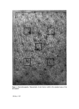
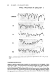
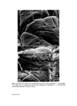
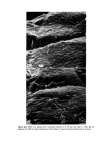
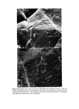
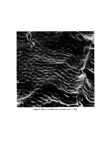
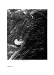
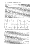
Emollients and the stratum corneum 623 after 5 h but some 'recovery' was apparent 8 h subsequent to treatment. Higher magni- fications (x 1000) of replica surfaces from areas treated with Emollient B indicated that a change in corneocyte morphology had taken place subsequent to treatment (Fig. 5a and b). The individual corneocytes often appeared thicker and the edges were more rounded and 'rolled' rather than angulated as in the controls. Figure 6a and b are scanning electron micrographs of replicas from both the untreated and the Emollient B treated legs of a woman with 'dry skin'. Although the emollient appears to have had a 'smoothing' effect on the surface furrows the uplifting of layers of surface corneocytes characteristic of dry skin (Nicholls, King and Marks, unpublished observations) were still apparent. No obvious changes were observed in scanning electron micrographs of SSBs from treated sites. In some specimens, however, ridge patterns appeared less accentuated at low magnifications. Figure 7 shows a typical ridge pattern of an SSB. DISCUSSION This study was designed to monitor the changes that take place at the skin surface and in the superficial parts of the horny layer following a single application of some of the commercially available emollients and bath oils. We have made no attempt at comparing the emollient preparations that we used although clearly comparative studies could easily be set up using the techniques that we describe. The results obtained suggested that the two emollients (oil in water emulsions) and the bath oil were all quite effective in 'smoothing' the SC. SEM of replicas from legs soaked in water only showed no obvious changes from the control site, although a signifi- cant decrease in surface contour was detected at 60 min post immersion. Other workers have detected little change in replica appearance after soaking for 1 h in warm water alone, although a clinical change was detected (4). Surface contour measurements of the hardened DPX replicas in the surfometer provided us with quantitative information about skin surface topography before and after treatment and demonstrated a flattening of contour after use of the materials. Since replicas were taken from adjacent sites (repeated replicas on one site may alter or remove some surface characteristic) the heterogeneity in surface architecture was reflected in the surface contour tracings. Subsequent to treatment, however, the reduction in surface contour was usually greater than normal site variation in each individual. These observed changes seemed to cor- relate with the morphological appearance of the specimen when examined by SEM, i.e. a filling in of the topographical 'valleys'. The small area and peak height changes demonstrable in SSB surfometer tracings suggested that some alteration in the internal structure of superficial corneum had also taken place subsequent to treatment although hardly any change could be demonstrated when the SSBs were inspected by SEM. Photography of skin sites has been used pre- viously as a subjective means of evaluating emollients (5). A final magnification of x 8 was used in this study. The consistent (but non-significant) increase in surface furrow width on our macrophotographs is difficult to explain, but may be due to an optical effect from the decreasing furrow depth after application of emollient or bath oil. We do not believe that this method has a great deal to offer and believe that more accurate data can be obtained by contour measurements of replicas. Furthermore, we suggest that it is particularly helpful when assessing 'skin surface active materials' to use a combination

624 S. Nicholls, C. S. King and R. Marks of replicas and SSBs for assessment. In this way it is possible to compare what is taking place at the surface with changes of some cell layers down in the SC. We hope to employ the techniques described in this study to evaluate the effects of chronic application of topical preparations thought to have a hydrating action on the SC and to correlate these effects with changes in the pattern of epidermal growth and maturation. ACKNOWLEDGMENTS We are-extremely grateful to Vick International Ltd, and Westwood Pharmaceuticals Inc for generous assistance towards these studies. REFERENCES 1 Marks, R. and Dawber, R. P. R. Skin surface biopsy: an improved technique for examination of the horny layer. Brit. J. Dermatol. 84 117 (1971). 2 Nicholls, S., King, C. S. and Marks, R. Morphological and quantitative assessment of physical changes in the horny layer in Ichthyoses. In: Marks, R. and Dykes, P. J. The Ichthyoses. MTP Press, Lancaster, U.K. (1978). 3 Marks, R. and Pearse, A.D. Surfometry: a method of evaluating the internal structure of the stratum corneum. Brit. J. Derrnatol. 92 651 (1975). 4 Garber, C. A. and Nightingale, C. T. Characterizing cosmetic effects and skin morphology by scanning electron microscopy. J. $oc. Cosmet. Chem. 27 509 (1976). 5 Quattrone, A. J. and Laden, K. Physical techniques for assessing skin moisturization. J. $oc. Cosmet. Chem. 27 607 (1976).
Purchased for the exclusive use of nofirst nolast (unknown)
From: SCC Media Library & Resource Center (library.scconline.org)