j. $oc. Cosmet. Chem., 29, 777-782 (December 1978) Effects of harvesting techniques on hydration dynamics: gravimetric studies of stratum corneum ROBERT L. RIETSCHEL and WILLIAM A. AKERS Department of Dermatology Research, Letterman Army Institute of Research, Presidio of San Francisco, CA 94129. Received October I I, 1977. Synopsis STRATUM CORNEUM specimens HARVESTED by several methods commonly used for in vitro studies have been compared by HYGROSCOPICITY. Cantharidin blisters give superior data when compared to heat, trypsin and ammonia fume separated specimens. As the cantharidin data represent skin from a different age group and body site, a comparison to animal and human data in the literature is made. INTRODUCTION Many experiments have been done using isolated sheets of stratum corneum. In 1963, Kligman and Christophers reported three techniques for preparation of these sheets: 1) exposure to ammonia fumes for 30 min in a closed vessel over concentrated ammo- nium hydroxide, 2) heat separation by immersing skin sandwiched between metal plates in a water bath at 56øC for 2 min followed by the removal of the epidermis, which is floated overnight in a buffered 0.0001% solution of trypsin and 3) in- traepidermal blistering with a 0.2% cantharidin solution occluded for 8-10 hr (1). They claimed that stratum corneum prepared by canthardin and heat methods was the same with respect to water diffusion and stress-strain characteristics, but no data was presented. Onken and Moyer obtained stratum corneum by digesting skin samples for 24 hr in 3% trypsin (2). Proteolytic enzymes other than trypsin have been used. Kligman and Christophers used papain, pepsin, ticin and elastase, but found them less effective than trypsin. Pronase is also useful (3). Middleton used 2M urea with 0.5% trypsin to obtain guinea pig foot-pad corneum which he compared with foot pad removed at 60øC for 30 min (4). He found no difference in the water binding ability of guinea pig foot pad prepared in these two ways. Another popular technique requires immersing skin in 2M sodium bromide for 2 hr at 37øC (5). Stretching has also been employed (6). Marzulli uses tape stripping of stratum corneum eluted with organic solvents (7). Each technique has its limitations. Some can only be utilized on excised skin. Several methods employ prolonged water immersion. The effects of enzymes on epidermal 777

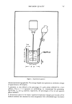
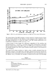
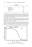
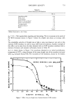
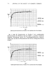
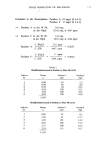
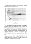
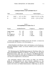
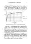
778 JOURNAL OF THE SOCIETY OF COSMETIC CHEMISTS protein cannot be ignored (8-9). Singer and Vinson showed a decrease in water binding capacity in corneum of neonatal rats after 17 hr of water immersion (10). At 93% rela- tive humidity they found water immersed corneum had a 47% increase over its dry weight as compared to 60% for the control. However Scheuplein reported that in comparing heat-separated stratum corneum to cantharidin blister tops, "neither tech- nique measurably damages the membrane. Some protein denaturation and dissolution must indeed occur but if this changed permeability, the differences are within the average experimental error, viz 20%, and cannot be isolated from the variations between different samples." (11) Polano et al. studied the effect of heat plus trypsin preparation of stratum corneum by measuring in vitro penetration of methyl nicotinate. They found no significant dif- ference in penetration utilizing 1-16 min immersions in 60øC water and concentra- tions of trypsin up to 0.1% (12). It seems reasonable that the stratum corneum is damaged by the separation procedures but due to difficulty in quantifying the denaturation, many investigators have chosen to ignore it. A sensitive, accurate and highly reproducible gravimetric method for studying the uptake of water vapor by stratum corneum samples from at- mospheres of controlled humidity and temperature has recently been developed (13). By studying hygroscopicity with this device, stratum corneum specimens prepared by several of the more commonly used methods described above have been compared. Anderson, Cassidy, Hansen et al. have recently shown that in vivo evaluation of dry skin correlates with in vitro hygroscopicity (14). Thus hygroscopicity seems a reason- able way to assess one factor in the normaIcy of stratum corneum samples. MATERIALS AND METHODS Abdominal skin from autopsy cases was collected. The specimens were refrigerated, but never frozen, until separation procedures could be effected. Each piece of skin was divided into four pieces. The first piece was separated by heat at 56øC for 3 min in a water bath. The second was separated as the first, then placed in 3% trypsin for 24.hr. The third was floated, epidermis down, in a 3% trypsin solution for 24 hr. (The 3% concentration was chosen to maximize any trypsin related effect.) And the fourth was removed by the ammonia fume method. All specimens were stored over a disiccant until ready for use. A 4-mm punch was taken from each specimen for comparison. The weight of each 4-mm piece was taken in a special chamber containing a Cahn elec- trobalance, controlled temperature and relative humidity regulated by saturated salt solutions (13). The per cent increased over dry weight of each sample was noted at 62 and 90% relative humidity and 20øC. Cantharidin blisters were prepared by placing a 2-cm by 2-cm piece of moistened filter paper containing 0.5 mL of a 0.2% cantharidin solution on the backs of volunteers for 3-4 hr. The filter paper was covered sequentially with Saran wrap © , Mystic tape © , Reston foam © and finally Micropore tape ©. After removing the occlusive dressing, the blister was allowed to fill overnight before removal of the top. Blister tops were stored over a desiccant and measurements were taken as above. RESULTS Separation techniques proved to be more variable than reported. Ammonia fumes did not always give good separation of dermis from epidermis. Trypsin was somewhat
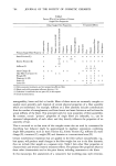
Purchased for the exclusive use of nofirst nolast (unknown)
From: SCC Media Library & Resource Center (library.scconline.org)