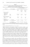
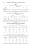
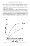
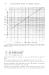
COMPARISON OF TOPICAL VEHICLES 305 FRANZ DIFFUSION CELLS (Crown Glass Co., Somervd/e, NJ) The cells are made up of the cell cap (donor) and the cell body (receiver). The cell cap was open to the air and allowed the application of a finite dose to the membrane. The volume of the cell body was 11 mL. The skin was mounted between the two ball joints, on an O-ring, using a pinch-type, ground joint clamp. The available area for diffusion was 3.14 cm 2. The epidermal side of the skin was exposed to ambient labo- ratory conditions, while the dermal side was bathed by a phosphate-buffered saline solution containing 0.02% thimerosal. Stirring of the receiver solution was accom- plished by a teflon-covered magnetic stirring bar, driven by an external magnet. The experiments were conducted at ambient temperatures (21 _+ IøC). PREPARATION OF SOLUTIONS The compositions of vehicles used in this study are given in Table II. An excessive amount of steroid was added to a 10 mL test tube containing the vehicle. The test tubes were shaken mechanically at room temperature for 24 hours. The saturated solutions were centrifuged (IEC HN-SII Centrifuge, International Equipment Co., Needham, MA) until the supernatant contained no crystals as determined by micro- scopic analysis. The crystal-free supernatant was then analyzed by HPLC for the con- centration of steroid. IN VITRO PERCUTANEOUS A•SORPTION STUDY Skin preparation and mounting. Mice were sacrificed by severing the spinal cord. A rectangular section of the abdominal skin was excised from the animal with surgical scissors. Adhering fat and other visceral debris were removed carefully from the under surface with tweezers. The skin was then mounted between the two cells and the receiver side was filled with the phosphate-buffered saline solution. Permeation. Following mounting, at time zero, 100 }xL of test solution was placed on the epidermal surface. One hundred [xL was sufficient to spread across the entire epi- dermal surface. Samples were withdrawn from the receiver compartments at 6, 24, 30, 48, and 72 hours and were analyzed by HPLC. Area under the curve was used to quantirate drug concentration in the receiver solution. Each experiment was performed at least in triplicate. The permeability data was plotted using the total amount of drug penetrated as a Table II Compositions of Vehicles Used for In Vitro and In Vivo Testing Parts by Volume Ingredient Vehicle No. 1 Vehicle No. 2 Sorbitan Stearate 1 1 Polysorbate 60 3 3 Propylene Glycol 10 -- Caprylic/Capric Triglyceride -- 10 Water 70 70

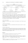
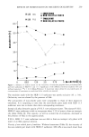
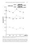
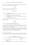
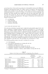
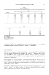
306 JOURNAL OF THE SOCIETY OF COSMETIC CHEMISTS function of time. Steady-state flux was often not observed until the third time period. The slope of the linear portion gave the total flux, JT. dM JT- dt' (1) where M = total amount of drug penetrated (mg). The steady-state flux JT can be described by: AKm CvD JT = , (2) h where A = the diffusional area (cm2). Km = the apparent partition coefficient between the stratum corneum and the vehicle. Cv = the drug concentration in the vehicle (mg/mL). D = the apparent diffusion coefficient of the drug through the stratum cor- neum (cm2/hr). h = the effective barrier thickness (cm). Higu•hi (31) expressed JT in terms of the thermodynamic activity of the penetrating agent in its vehicle: AD a v JT = -- , (3) •/s h where a• = the activity of the drug in the vehicle. •/s = the activity coefficient of the drug in the barrier. The values of •/s, A, D, and h are constant unless the vehicle alters the barrier. When the barrier is stable, changes in flux may be attributed to the changes of the thermo- dynamic activity of the drug in the vehicle. In saturated solutions, the thermodynamic activity is determined by the crystalline state of the drug and theoretically is the same from vehicle to vehicle. Thus, according to equation 3, the flux from the saturated solutions should be the same, even when the actual concentration of the steroid in the vehicles is different. IN VIVO VASOCONSTRICTION STUDY The concentrations of steroids used in this study are shown in Table III (concentrations were at or above saturation, viz., as solutions or slurries). The influence of the vehicle Table III Steroid Concentration Used in Vasoconstriction Assay Concentration Steroid (mg/mL) Hydrocortisone 17-butyrate 1.0 Desonide 0.5 Difiorasone Diacetate 0.5
Purchased for the exclusive use of nofirst nolast (unknown)
From: SCC Media Library & Resource Center (library.scconline.org)