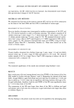
IN VITRO EFFECT OF WHITENING COSMETICS 363 melanocytes were cultured for about two weeks in MCDB153 medium containing 0.13 mM CaCI 2, 10 ng/ml epidermal growth factor (EGF), 5 •g/ml insulin, 0.5 •g/ml hydrocortisone, 1 ng/ml recombinant basic fibroblast growth factor (rbFGF), 10 ng/ml phorbol 12-myristate, 13-acetate (PMA) and 0.2% v/v of bovine pituitary extract. After replacement of the medium with MCDB 153 without rbFGF and PMA, the cells were cultured for a further two days. The cultured melanocytes were placed in 96-well plates at a density of 12,500 cells/cm 2. Two plates, one for the measurement of tyrosinase activity and the other for the measurement of cell viability, were cultured for one day. Hydroquinone and linoleic acid at final concentrations of 0.001, 0.005, 0.01, and 0.05 mM, and arbutin, kojic acid, and ascorbic acid at final concentrations of 0.01, 0.05, 0.10, 0.50, 1.0, and 5.0 mM, were added to triplicate wells and cultured at 37øC for three days. CELL VIABILITY ASSAY The cell viability in the presence of each agent was evaluated by the MTT test (13). After culture, phosphate buffer (pH 7.4) containing 5 mg/ml MTT (3-(4,5-dimethylthiazol- 2-yl)-2,5-diphenyl tetrasolium bromide) was added to each well. The plate was incu- bated at 37øC for 4 hours, after which isopropanol containing 0.04 N HC1 was added. Viable cells formed dark blue formazan by cleaving MTT with mitochondria. After 30 minutes, absorbance was measured at 570 nm using 655 nm as a reference. To eliminate agent interference in the measurement, wells containing each test agent alone were incubated and reacted with MTT. Cell survival was calculated from the absorbance. TYROSINASE ACTIVITY ASSAY The cells were washed with PBS and lysed with 45 •1 of 1% Triton-X/PBS. After vibration, 5 •1 of 10 mM L-DOPA was added to the wells. After incubation of the plates at 37øC for 60 min, absorbance was measured at 475 nm in a Model-3550 ELISA Reader (Bio-Rad Lab., Richmond, CA). The absorbance values were compared with a standard curve obtained with mushroom tyrosinase (Sigma Chemical Co., St. Louis, MO) the standard curve was linear within the range of experimental values. The coefficient of correlation was determined as 0.999. MELANIN ASSAY Melanin content was determined according to the method described by Oikawa (14), which we modified. Briefly, human melanocytes (40,000 cells/well) cultured in 12-well plates in 1.0 ml of medium were cultured with arbutin (0.5 mM), kojic acid (0.5 mM), and ascorbic acid (0.5 mM) at 37øC for three days. The culture solution was aspirated, and 1.0 ml of 2.5% trypsin solution was added to each well. The cells were detached, placed in an Eppendorf tube, and centrifuged at 1,000 rpm in a Hitachi CR 15 refrigerated centrifuge to obtain cell pellets. The pellets were mixed with 5% trichlo- roacetic acid, agitated well, and centrifuged at 10,000 rpm to deposit melanin. The melanin sediment was washed with PBS and mixed with 1N NaOH for dissolution, and the absorbance at 475 nm was measured. The amount of melanin per cell was calculated

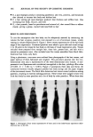
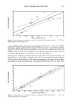
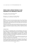
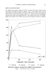
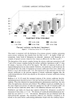
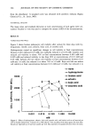
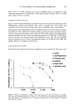
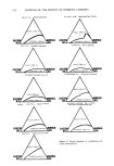
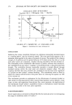
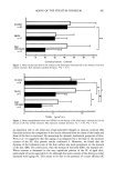
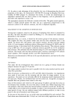
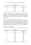
364 JOURNAL OF THE SOCIETY OF COSMETIC CHEMISTS from the absorbance. A standard curve was obtained with synthetic melanin (Sigma Chemical Co., St. Louis, MO). STATISTICAL ANALYSIS The mean value and standard deviation at each concentration of each agent were cal- culated. Student's t-test was used to compare the means (-+SD) of the determinations. RESULTS VIABLE CELLS PER WELL Figure 2 shows human melanocyte cell viability after culture for three days with hy- droquinone, linoleic acid, arbutin, kojic acid, or ascorbic acid. Hydroquinone caused no significant changes in cell viability at final concentrations between 0.00! mM and 0.0! mM, but markedly reduced it at 0.05 mM. Linoleic acid reduced cell viability to about 70% at final concentrations between 0.00! mM and 0.005 mM and reduced viability to less than 20% at concentrations of 0.01 mM and 0.05 mM. Arbutin did not reduce cell viability at final concentrations between 0.0 ! mM and 1.0 mM, but reduced it to about 74% at 5.0 mM. Kojic acid did not reduce cell viability at final concentrations between 0.0! mM and 1.0 mM, but reduced it to 3000 2000 lOOO o.ool o none --- hydroquinone -- linoleic acid -e-arbutin • kojic acid [] ascorbic acid ß . . ....al ß , , .an..I ß . ß nl=..I ß ß . 0.010 0.100 1.000 10.000 rnM Figure 2. Effects of hydroquinone, arbutin, kojic acid, ascorbic acid, and linoleic acid on cell survival per well in human melanocytes. Cultures of 12,500 cells/cm 2 were incubated with these agents for three days. Results are expressed as cell survival number per well. Bars represent of standard deviation from the mean.
Purchased for the exclusive use of nofirst nolast (unknown)
From: SCC Media Library & Resource Center (library.scconline.org)