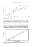
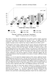
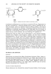
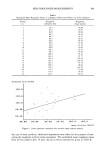
j. Soc. Cosmet. Chem., 42, 385-391 (November/December 1991) Comparative studies of skin roughness measurements by image analysis and several in vivo skin testing methods KARLHEINZ SCHRADER and STEPHAN BIELFELDT, Creachem GmbH, Research Institute for the Cosmetics Industry, Max-Planck-Stra?e 6, D-3450 Holzminden, Germany. Received October 24, 1989. Synopsis Measurement of skin roughness on the volar forearm in humans using image analysis correlates significantly with skin moisture assessment by measuring the dielectric constant of skin (r = 0.57), skin roughness measurement by methylene blue adsorption (r = 0.48), and roughness measurement using a stylus instrument (0.32). This study supports the conclusion that the described image analysis method is useful in the evaluation of skin roughness. INTRODUCTION Skin roughness is a very important parameter in the characterization of cosmetic skin properties. Smooth, supple skin is a perceptible effect that can be achieved by means of cosmetic skin care. In the past twenty years many procedures have been developed to obtain direct or indirect information about skin roughness. Some methods, such as measurement of the skin's moisture content, are frequently used because a close correlation with skin rough- ness exists. In this study, an image analysis method of evaluating skin profile impressions was correlated with three other recognized procedures: moisture content by dielectric con- stant measurement, skin roughness measurement by methylene blue adsorption, and roughness measurement by profilometry. MATERIALS AND METHODS MEASURING SKIN ROUGHNESS WITH AN IMAGE ANALYSIS SYSTEM Evaluation of skin impressions by image analysis has been described principally by 385

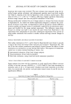
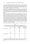
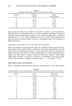
386 JOURNAL OF THE SOCIETY OF COSMETIC CHEMISTS Corcuff et al. (1-4). The roughness of the profile is determined by measuring the shadows produced on the impression by means of an oblique light source. For this study, negative impressions from the volar forearm were measured. A colorless and pigment-free silicon mass was used as the material for taking the impressions. The impressions were evaluated at approximately 20 x magnification under the micro- scope. They were lit from below by a fiber optic light source at 45 ø to the perpendicular axis. The skin relief, which appears three-dimensional, was taken by a video camera and stored for further image processing in the image analyser (Quantitative Image Analyser, Carl Zeiss, D-2000 Hamburg 1, Germany) of the image analysis system. A grey level discrimination creates a binary image. The white phase consists of the illuminated "slopes" (negative furrows). Other structures are excluded. To eliminate further insig- nificant parts of the image, two different algorithms were used. All these objects in the image with the longer axis direction not perpendicular to the incident light direction were eliminated. An angle mismatch of not more than 30 ø was accepted. Also objects with a relation feret y/feret x less than 2 were eliminated. The direction y is perpendicular to the incident light direction. This was done to eliminate air bubbles and round particles in the measurement. The width of the correctly shaped and orientated "slopes" were measured by a cordlength algorithm all over the image. To get data of all furrows independent of the orientation, the replica was turned in steps of 30 ø and measured in each direction of 0 ø to 180 ø . The mean of all six directions (an average of about 1.500 individual measure- ments) was used as the roughness value of one replica. MEASURING SKIN ROUGHNESS WITH A STYLUS INSTRUMENT In the past 10 years, skin roughness measurement using stylus instruments has been largely perfected and automated. The various measuring and evaluation procedures are well-documented (references 5-8), and we need not examine them further here. In the methods used for this study, silicon impressions of the volar forearm were prepared. A variety of dental masses are suitable materials for making the impressions. Xantopren © from Bayer AG, Leverkusen, was used in this study. The impressions were evaluated by means of a Hommel T 20 © stylus unit (Hommel- werke GmbH, D-7730 VS-Schwenningen, Germany) with fully automatic sample feed. Circular © silicon impressions (skin negatives) were sectioned off radially in 12 different directions. The scan length was 12.5 mm. The sensor has a tip radius of 5 •m. The parameter R z was measured, and in order to determine it, the scan length was divided into five equal-sized areas. In each area, the distance from the highest peak to the lowest trough was established. The average of these five distances is R z (2). The mean of the 12 values for R z was taken for each impression. MEASURING THE SKIN MOISTURE The moisture of the skin in vivo can be determined by various methods. The most widely used procedures are infrared spectroscopy, the measurement of electrical properties, photoacoustic spectroscopy, and the determination of transepidermal loss of moisture
Purchased for the exclusive use of nofirst nolast (unknown)
From: SCC Media Library & Resource Center (library.scconline.org)