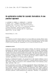
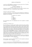
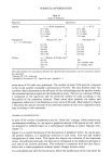
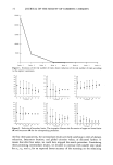
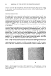
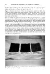
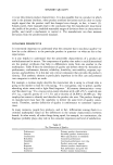
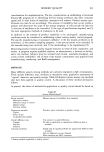
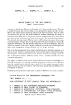
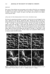
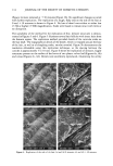
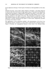
REPLICATION OF SKIN 113 RESULTS AND DISCUSSION The effects of casting the positive replica from the same Reprosil mold at 0, 77, and 81 days are shown in Figure 2. The overall surface topography was not observed to be affected at either low (x 50) or higher magnification (x 250) at 77 days after the sample was taken (Figure 2a,b,d,e). The effect of repeated use of the same Reprosil mold is illustrated in Figures 2c and 2f. Surface pitting (P) was evident with repeated use however, in practice the Reprosil molds are only used once. The globular structures (G) were reproduced accurately, except in Figure 2d, where one of the structures was absent. The globular structures are fragile and may easily be lost either during removal of the Reprosil negative from the epoxy or during the final mounting of the sample. Figure 3 shows the effects of replicating the skin at 30-minute intervals at a magnifi- cation of x 150. Several loose flakes of skin that were present in the initial replica / 881469 10KV X150 •00u• 8-5)8 Figure 3. Replication of the same area of the forearm at 30-minute intervals: (a) 0 minutes (b) + 30 minutes (c) + 60 minutes (d) + 90 minutes.

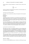
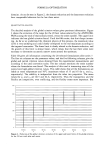
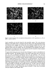
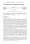
114 JOURNAL OF THE SOCIETY OF COSMETIC CHEMISTS (Figure 3a) were removed at q- 30 minutes (Figure 3b). No significant changes occurred with further replication. The replication of a rough, flaky area on the sole of the foot at 0 and q- 30 minutes is shown in Figure 4. No loss of detail was evident at either low (x 50) or higher (x 500) magnification. Scales were found to remain intact with little or no disruption. The suitability of the method for the replication of fine, delicate structures is demon- strated in Figures 5 and 6. Figure 5 illustrates several hair follicles with intact hairs from the forearm region. The replication method provided details of the cuticular scales on the hair shaft. The topographical details of the sheath, which is wrapped around the base of the hair, as well as of sloughing scales, are also revealed. Figure 5b demonstrates the resolution obtainable using this replication technique, as the spacing between the cuticles is approximately 5 to 10 mm. Figure 6 shows the replication of intricate, fragile structures present on the surface of the leaves of two plants: velvet leaf (Figures 6a, 6b) and coleus (Figures 6c, 6d). Details were excellently reproduced, illustrating the utility •$•IPL ' , 'LE • SOLE.. ß -. ., ß Figure 4. Replication of the sole of the foot: (a) and (b) 0 minutes (c) and (d) + 30 minutes.
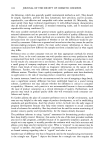
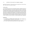
Purchased for the exclusive use of nofirst nolast (unknown)
From: SCC Media Library & Resource Center (library.scconline.org)