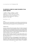
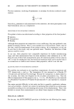
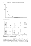
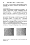
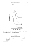
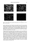
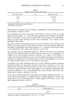
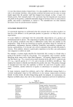
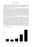
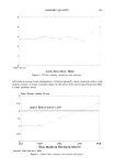
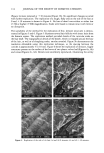
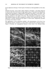
REPLICATION OF SKIN 117 •0295.S 10KY X500 60um SAMPLE'#?9-'': FORE Figure 8. Topographical features of three areas of the forehead, before treatment with either facial toner or facial lotion. '$AMP•E• • "- •QJJP •l .#.5.9 TDk0mSn) ' ' ' _. ' '- c•_ .-, '•.. d , .-•-•. ,. ,..• •' •, t•••:•..%•g•Tk •- ._•,--'• , •(•:lhr• • "•' PgA'" ' :3h•)'- . S,MPkEJ: :'H Figure 9. Topographical features of three are• of the forehead after application of a facial lotion: (A), (C), and (E) ten minutes after application (B) at one hour (D) at three hours (F) at five hours after application. treatment with either the facial toner or facial lotion (a blank). The same areas of the forehead are shown in Figure 9, 10 minutes after application of the facial lotion (A, C, E) and at one hour (B), three hours (D), and five hours (F) after application of the facial lotion. A substantial amount of the polymer was found on the forehead at one hour after

118 JOURNAL OF THE SOCIETY OF COSMETIC CHEMISTS application of the product. At three and five hours after application of the facial lotion, only a small amount of the polymer remained. CONCLUSIONS A two-step replication process that utilizes Reprosil and Spurr's embedding medium has been critically evaluated for use in the study of personal care products on human skin. The high dimensional stability of the Reprosil material makes collection of entire data sets possible. This is especially convenient in multiple-person studies, where all samples can be collected and processed together under similar conditions at a later date or be sent collectively to an independent testing laboratory. The replication process is particularily well suited for the replication of fine, delicate structures. Details of hair follicles and the surface of leaves were excellently reproduced. The replication process may be easily and conveniently applied to the study of effects of personal care products on human stratum corneum, as long as the identical testing site is utilized for before and after comparisons. REFERENCES (1) C. A. Garber and C. T. Nightingale, Characterizing cosmetic effects and skin morphology by scan- ning electron microscopy, J. Invest. Dermatol., 27, 509-531 (1976). (2) C. H. Pameijer, Replication techniques with new dental impression materials in combination with different negative impression materials, Scanning Electron Microsc., II, 571-574 (1979). (3) M. A. Hayat, Transmission Electron Microscopy (Academic Press, San Diego, 1986), pp. 6-7.
Purchased for the exclusive use of nofirst nolast (unknown)
From: SCC Media Library & Resource Center (library.scconline.org)