202 JOURNAL OF THE SOCIETY OF COSMETIC CHEMISTS (9) J. W. Jorgenson and K. D. Lukacs, High-resolution separations based on electrophoresis and elec- troosmosis, J. Chromatogr., 218, 209-216 (1981). (10) J. W. Jorgenson and K. D. Lukacs, Capillary zone electrophoresis, Science, 222, 266-272 (1983). (11) S. Terabe, K. Otsuka, K. Ichikawa, A. Tsuchiya, and T. Ando, Electrokinetic separations with micellar solutions and open-tubular capillaries, Anal. Chem., 56, 111-113 (1984). (12) S. Terabe, K. Otsuka, and T. Ando, Electrokinetic chromatography with miceliar solution and open-tubular capillary, Anal. Chem., 57, 834-841 (1985). (13) R. J. Nelson, A. Paulus, A. S. Cohen, A. Guttman, and B. L. Karger, Use ofpeltier thermoelectric devices to control column temperature in high-performance capillary electrophoresis, J. Chromatogr., 480, 111-127 (1989).

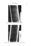
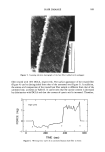
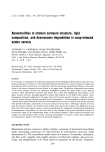
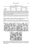
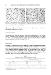
j. Soc. Cosmet. Chem., 45, 203-220 (July/August 1994) Abnormalities in stratum corneum structure, lipid composition, and desmosome degradation in soap-induced winter xerosis ANTHONY V. RAWLINGS, ALLAN WATKINSON, JULIA ROGERS, ANA-MARIA MAYO, JAMES HOPE, and IAN R. SCOTT, Unilever Research, Edgewater, NJ (A.V.R., I.R.S.), and Unilever Research, Sharnbrook, Bedford, UK (A.W., J.R. , A.-M.M. , J.H. ). Received April 20, 1994. Presented in part at the 17th IFSCC International Congress, October 1992, and at the the Society for Investigative Dermatology, April 1993. Synopsis In an attempt to understand the underlying biochemical and morphological abnormalities that lead to the physical appearance of xerosis, we have examined lipids and desmosomes in stratum corneum of normal and soap-induced winter xerotic skin. In normal skin, electron microscopy revealed lipid bilayers in the lower layers of the stratum comeurn that were absent in the upper layers. In addition, desmosomes were present in the lower stratum comeurn but underwent degradation towards the upper surface of the stratum corneum. These observations contrasted with xerotic skin, which had disorganized lipid bilayers in the upper stratum corneum, although apparently normal lipid bilayers in the deeper tissue regions. Also, desmosomes remained undegraded in the upper layers of the xerotic stratum corneum, a finding corrobo- rated by western blotting showing increased levels of desmoglein 1. Chromatographic analysis of stratum comeurn lipids showed decreased ceramide and increased fatty acid levels in subjects with xerosis compared with normal individuals, particularly in the outer stratum corneum layers. Although ceramides were lost from the stratum comeurn, the increased levels of fatty acids may be due in part to the deposition of soap fatty acids. Our results support previous studies demonstrating the importance of desmosomal degradation in desquamation. Furthermore, we have been able to show changes in the normal membrane structure of intracellular lipids in the desquamating layers of the stratum comeurn. These studies also provide new insights into soap-induced winter xerosis, revealing abnormalities in stratum comeurn lipid composition and organization together with reduced desmosomal degradation. INTRODUCTION Mammalian stratum corneum is highly complex, consisting of specialized intracellular lipids surrounding corneocytes (1) interconnected by proteinaceous structures called desmosomes (2). The intercellular lipids consist of a mixture of ceramides, cholesterol, and fatty acids, together with smaller amounts of cholesterol sulphate, glucosyl cera- mides, and phospholipids (3,4). These lipids are important for the barrier, desquama- 203
Purchased for the exclusive use of nofirst nolast (unknown)
From: SCC Media Library & Resource Center (library.scconline.org)