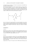
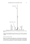
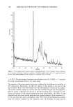
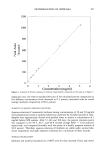
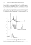
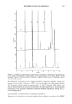
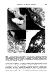
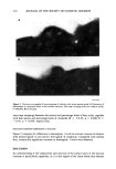
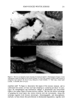
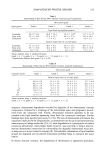
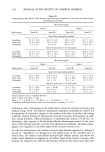
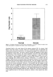
204 JOURNAL OF THE SOCIETY OF COSMETIC CHEMISTS tory, and mechanical properties of the stratum corneum (for review, see reference 5). Functionality is probably related to the capacity of stratum corneum lipids to form multiple lipid bilayers and their associated gel and liquid crystalline properties. During the normal maturation of the stratum corneum, individual cells are lost from its surface in a process called desquamation (6). For desquamation to proceed, the degra- dation of all the cohesive elements holding the cells together in the stratum corneum must occur. Stratum corneum lipids (1), lectins (7), and desmosomes (2,8) are thought to play roles in intercorneocyte cohesion. Recent findings, however, have indicated that desmosomes may be the main intercellular linkages in this tissue (2,8). Although initially thought to be non-functional within the stratum corneum due to their degra- dation in the lower layers (9, 10), studies have shown that some desmosomes, especially those associated with corneocyte interdigitations, persist intact up to the peripheral layers of the tissue (2, 11, 12). Supporting biochemical data has been provided by the persistence of desmoglein 1 (dsg 1) throughout the stratum corneum up to the peripheral layers where degradation appears to occur (11, 13). Thus, desmosomal degradation is believed to be an important part of the desquamatory process. Indeed, the enzymatic degradation of desmosomes has recently been shown to aid desquamation in palmoplan- tar and non-palmoplantar stratum corneum (12,14). Degradation of desmosomes is believed to be the result of the action of serine proteases (12,14-18), especially chy- tootrypsin-like (16) and possibly trypsin-like proteases (19). In addition to desmosomal proteins, the stratum corneum lectin, desquamin (20), may also play a role in intercorneocyte cohesion, although this has yet to be fully determined. Stratum corneum lipids, however, influence stratum corneum integrity, and hence their degradation is an important aspect of desquamation (1). Phospholipid and glucosylce- ramide hydrolysis occurs during the stratum compatum to stratum disjunctum transi- tion (3), and cholesterol sulphate hydrolysis is intimately associated with the loss of surface corneocytes (21,22). The most common problem affecting the stratum corneum is the accumulation of visible scales or corneocyte clumps on the surface of the skin, the severity of which can range from the genetic ichthyoses affecting a minority of the population (23,24), to xerosis, which concerns a large proportion of the population, particularly the elderly (25). Although the precise causes of skin xerosis are likely to be multifactorial (26), the resultant scaling is probably a consequence of perturbed degradation of the corneocyte adhesive elements, leading to aberrant desquamation. However, the relative contribu- tion of lipids and desmosomes to the disordered desquamatory process is poorly under- stood. Stratum corneum lipids have been well studied in xerosis, although no consensus in the role of lipids have been produced. Solvent (27) or surfactant (28) induced dry scaly skin was associated with intercellular lipid depletion. In contrast to these studies, the levels of stratum corneum fatty acids are increased, but ceramides, cholesterol, and cholesterol sulphate are reported not to be changed in subjects with winter xerosis (29). In addition, using electron microscopy, Fartasch et al. (30) did not find any abnormal- ities in stratum corneum lipid structure following surfactant use. An alternative view is that alterations in stratum corneum lipid composition, but not total lipid levels, result in skin scaling. In support of this, changes in lipid composition, especially ceramide subtypes, have been induced following surfactant treatment or tape stripping, and scaling has been elicited (31,32).

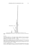
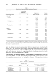
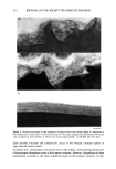
SOAP-INDUCED WINTER XEROSIS 205 The present study was designed to clarify the biochemical and morphological aberrations in stratum corneum lipids and desmosomes in skin xerosis. We investigated these components in particular because of the lack of consensus on the role of lipids in xerosis and because of the increased recognition of the role of desmosomes in stratum corneum cohesion and desquamation. The results demonstrate that stratum corneum ceramides are depleted in soap-induced winter xerosis however, there are no alterations in the relative ceramide sub-species. Additionally, we have shown that in soap-induced winter xerosis there was abnormal desmosomal degradation resulting in retention of these adhesive structures in the peripheral layers of the stratum corneum to produce skin scaling. MATERIALS AND METHODS SUBJECTS AND TREATMENTS Female Caucasians aged 30-40 years participated in this study. Subjects with genetic disorders or a history of atopy were excluded from the study (33), and all had normal skin at the start of the study. Normal controls were obtained from similar subjects not in the trial. To generate xerosis, all subjects refrained from the use of moisturizers and used only soap for cleansing three times per day for one week, as previously described (33), prior to grading and stratum corneum sampling. At the end of this period, the clinical severity of the xerosis of the dorsal surface of their hands was graded according to the following criteria: Grade 1: Normal skin. Grade 2: Mild xerosis characterized by small flakes of dry skin and whitening of dermatoglyphic triangles. Grade 3: Moderate xerosis, small dry flakes giving a light powdery appearance to the hand. Corners of dermatoglyphic triangles have started to uplift. Grade 4: Well-defined xerosis the entire length of a number of dermatoglyphic trian- gles have uplifted to generate large, dry skin flakes. Roughness is very evi- dent. STRATUM CORNEUM SAMPLINGS Skin biopsies were taken from the back of human hands using adhesive tape (Sellotape, DRG Products, UK). A 2 x 3-cm piece of tape was applied to the hands with gentle pressure by 20 strokes of a gloved finger and carefully removed. This procedure was repeated up to eight times on the same site, allowing analysis of the total stratum corneum sampled and stratum corneum of different depths the first tape was used to analyze the "outer stratum corneum" and the remainder pooled for the analysis of the "inner stratum corneum." Using electron microscopy techniques, it was found that approximately five layers of stratum corneum were removed during this process. Tape strippings were stored at -20øC until required. Corneocytes were detached from the tape in methanol with sonication, the tape was discarded, and the corneocytes together with methanol were dried under a stream of nitrogen.
Purchased for the exclusive use of nofirst nolast (unknown)
From: SCC Media Library & Resource Center (library.scconline.org)