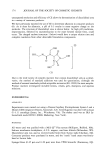
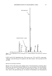
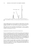
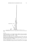
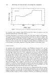
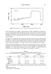
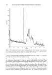
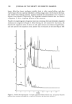
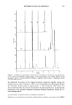
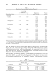
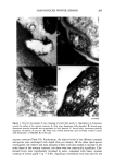
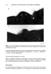
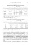
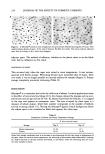
216 JOURNAL OF THE SOCIETY OF COSMETIC CHEMISTS In parallel with the morphological changes in the lipid bilayers, changes in stratum corneum lipid composition are also associated with winter xerosis. Our data demon- strated a decrease in the content of ceramide in xerotic skin comparison of inner and outer stratum corneum revealed that in the milder xerotic skin the decrease in ceramide levels was a surface phenomenon that extended to the deeper layers of the stratum corneum in the more severe xerotic grades. Interestingly, the ratio of the ceramide sub-species did not alter. Similar decreases in ceramides have been found in atopic dermatitis (48-50). Although others have shown increases in fatty acid levels in the stratum corneum of xerotic subjects (29), in our study the fatty acids only tended to increase. In both of these studies the increase in fatty acid levels in the stratum corneum may be due to deposition of soap-derived fatty acids. The reasons for the aberration in stratum corneum lipid structure in soap-induced winter xerosis are unknown, but they are probably related to diminishing ceramide and in- creasing fatty acid levels, which may lead to an alteration in the phase behavior of the stratum corneum lipids and result in crystallization of the lipid matrix. Friberg and Kayali (51) have demonstrated that saturated fatty acids offer little resistance to water transport their increased levels in the upper stratum corneum may influence lipid structure and barrier function. Although the levels of fatty acids do not show statistical differences in their concentrations between the inner and outer layers of the stratum corneum due to the large inter-individual variation, their mass levels were nearly doubled in subjects with xerosis. The fatty acids may be derived from the soap for bathing, due to the hydrolysis of ceramides by a ceramidase (52), or may be of sebae- ceous origin. Excess fatty acids have been found in the lipid fractions derived from low-humidity-induced dry skin samples of pigs (53), indicating intrinsic origins rather than extraneous sources. It is therefore possible that the alteration of the ratio of the three major lipid components, fatty acids, steroIs, and ceramides, causes phase separa- tion of lipids at the surface of the stratum corneum. The excess fatty acid levels may further exacerbate the structural defects of the intercellular lipid fatty acids alter the phase properties of phospholipid bilayers (54). Such phase separations have also been seen with lovastatin-treated animals (55). Whether the surface abberations in lipid structure and composition together with degradation in xerotic desmosomes are linked is presently unknown. However, it is possible that changes in lipid structure or composition could interfere with the enzymes responsible for desmosomal degradation. To date, one candidate as a desmosome- degrading desquamatory enzyme is stratum corneum chymotryptic enzyme (SCCE) (16). It is possible that this, or similar desquamatory hydrolases (19), could be inhibited by the altered lipid environment or denatured by direct soap action, or that, alternatively, these enzymes could be affected by the external environment, causing either denatur- ation of the protein or prevention of access to the desmosome. It is interesting in this respect that other chymotryptic enzymes are inhibited by fatty acids (56) and that we, and others (29), find increased fatty acid levels in soap-induced winter xerosis. Another possible linkage between the desmosome and lipid elements in skin xerosis could be at the level of the lameliar bodies in the granular layer. These lysosome-like structures are the source of the stratum corneum intercellular lipids, and possibly also of the desquamatory enzymes (3,42). Environmental effects could feed back to the granular layer, affecting delivery of their contents. However, it is difficult to see how

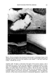
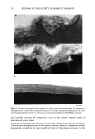
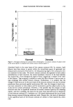
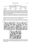
SOAP-INDUCED WINTER XEROSIS 217 such a mechanism would manifest itself in the surface layers of the stratum corneum after only one week of insult to the stratum corneum, which is insufficient time for full stratum corneum turnover, suggesting that the desmosomal and lipid abnormalities we see in the present study are a result of environmental insult to the surface layers of the stratum corneum. It is possible, however, that this mechanism can operate in other more chronic disorders (31,32,42,49). Desmosome retention, however, was also associated with structural abnormalities in stratum corneum lipid lamellae, especially in the outer tape strippings of xerotic skin compared with normal skin. Nevertheless, the gross aberrations in lipid structure were in areas of the tissue where desmosomes were not present and not in the regions where desmosomes were present. The close opposition of corneocytes due to the desmosomal linkages may help to prevent disruption of lipid structure in these parts of the stratum corneum. Although desmosomes largely influence corneocyte cohesion (8), changes in the physical properties of the stratum corneum lipids will also probably influence intercorneocyte cohesion properties and thereby influence desquamation. Changes in the physical properties of stratum corneum lipids are reported to occur in the different layers of the stratum corneum (57). The relationships of these structural changes and skin xerosis remain to be determined. Nevertheless, it has been reported that skin extensi- bility decreases with increasing severity of soap-induced winter xerosis (58). Although we suspect that the diminished elasticity of the stratum corneum arises as a result of increased numbers of desmosomes, changes in lipid structure and reduced natural mois- turizing factors (59) will also contribute. Over three decades ago Kligman (60) suggested that cell cohesion in the stratum corneum was dependent upon an "intercellular cement" that is predicted to become less stable near the surface of the skin or to be degraded by enzymes. We and others (2,12,39,42) have now shown that desmosomal degradation is an important event leading to desquamation. However, we have also provided some morphological evidence for the breakdown of lipid bilayers in the surface layers of the stratum corneum in normal skin. In addition, we also show for the first time changes in the morphology of lipid bilayers and the persistence of desmosomes in severe xerosis. In summary, we have used tape stripping to analyze the roles of lipids and desmosomes in the peripheral layers of the stratum corneum in normal and soap-induced winter xerotic skin. Our results showed that in normal skin there is desmosomal retention close to the surface of the stratum corneum, where their final degradation occurs. Similarily, degradation or disruption of the stratum corneum multiple lipid bilayers apparently occurred at the tissue periphery. The aberrations in soap-induced winter xerosis also appeared to manifest themselves in the upper layers of the stratum corneum, with retention of desmosomes and a collapse of the bilayer structure to give a disorganized lipid matrix of completely different structure to that of normal skin. The resultant interference in the breakdown of the intercorneocyte cohesive forces at the stratum corneum surface appears to account for the scaling that is the major symptom of soap-induced winter xerosis. ACKNOWLEDGMENTS We would like to thank Dr. T. Egelrud, Department of Dermatology, UMEA, Sweden,
Purchased for the exclusive use of nofirst nolast (unknown)
From: SCC Media Library & Resource Center (library.scconline.org)