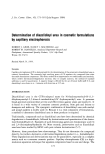
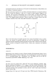
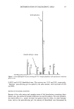
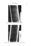
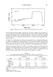
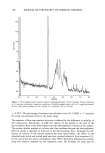
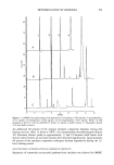
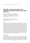
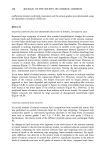
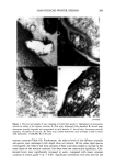
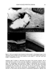
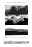
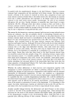
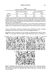
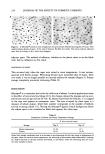
212 JOURNAL OF THE SOCIETY OF COSMETIC CHEMISTS Figure 4. Electron micrograph of tape strippings of subjects with severe xerosis (grade 4). Aberration in lipid organization toward surface of stratum corneum: A. First strip disorganized lipid lamellae. B. Second strip disorganized lipid lamellae. C. Third strip normal lipid lamellae. (x200,000 Bar 0.05 lipid lamellae structure and composition, occur at the stratum corneum surface in soap-induced winter xerosis. In normal skin, desmosomes were found close to the surface, demonstrating persistence of desmosomes throughout most of the stratum corneum. However, degradation of these desmosomes occurred in the most superficial layers of the stratum corneum. In this

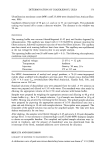
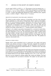
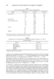
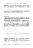
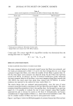
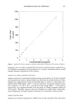
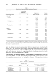
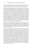
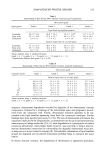
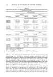
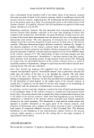
SOAP-INDUCED WINTER XEROSIS 213 Table I Relationship of Skin Xerosis With Stratum Corneum Lipid Composition Skin xerosis grade Lipid species Grade 1 Grade 2 Grade 3 Grade 4 Lipid levels (ng lipid/lxg protein) Ceramides 64.9 +- 34.4 68.6 -+ 30.4 39.2 +- 14.9' 37.5 +- 14.1' Fatty acids 62.1 -+ 34.6 67.4 -+ 32.7 60.5 +- 37.0 54.9 +- 28.1 Cholesterol 3.9 +- 2.1 7.7 -+ 4.2 4.4 -+ 2.0 4.6 -+ 2.3 Relative lipid levels (% of total lipids) Ceramides 47.1 +- 17.4 48.3 -+ 8.6 40.2 + 13.2 38.3 +- 11.2 Fatty acids 49.7 + 18.6 46.2 + 9.8 55.0 + 12.0 56.0 + 10.8 Cholesterol 2.0 -+ 1.9 5.5 +- 2.6 4.8 + 2.4 5.2 + 3.2 Values represent mean +-- standard deviation. Grade 1, n = 8 grade 2, n = 8 grade 3, n = 12' grade 4, n = 12. * Significantly different from grade 1 (p 0.05). Table II Relationship of Skin Xerosis With Stratum Corneum Ceramide Composition Skin xerosis grade Ceramide species Grade 1 Grade 2 Grade 3 Grade 4 Relative lipid levels (% of total lipids) 1 10.6 +__ 2.8 12.4 + 3.6 15.6 + 3.3 15.7 --- 8.4 2 20.2 + 4.2 23.2 + 3.8 24.6 + 3.8 28.6 ___ 6.1 3 18.0 _+ 2.9 17.5 +-- 3.2 17.7 +-- 4.5 16.1 +__ 4.5 3a 10.6 +-- 4.6 6.4 --- 1.5 6.7 --- 3.3 6.7 --- 2.7 4/5 21.8 --- 5.1 22.9 +-- 1.5 20.9 --- 2.5 19.7 -+ 5.4 61 7.0 -+ 3.5 6.0 +-- 1.7 3.7 +-- 2.2 2.4 +-- 1.6 611 12.4 --- 3.1 11.7 --- 1.4 10.4 --- 2.6 9.5 --- 2.7 Values represent mean +--- standard deviation. Grade 1, n = 8 grade 2, n = 8' grade 3, n = 12' grade 4, n = 12. sequence, desmosomal degradation occurred by digestion of the desmosomes' internal components, followed by a widening of the intercellular space and progressive detach- ment from the corneocyte wall. Finally, desmosomal remnants appeared to be sur- rounded with lipid lamellae separating them from the corneocyte envelopes. Similar findings have been reported previously (12,39). The loss of desmosomal attachment has important implications for desquamation, as desmosomes are the principal proteinaceous intercorneocyte linkage molecules (2,8,11) their degradation is a prerequisite to surface corneocyte detachment. Intercellular lipids, however, may also help to reduce intercor- neocyte desmosomal interactions by surrounding the degraded desmosomal structures, or may even act as anti-cohesive elements (8). Nevertheless, reformation of lipid lamellae around the degraded and vacuolated desmosome probably helps to maintain the water barrier. In xerotic stratum corneum, the degradation of desmosomes is apparently perturbed,
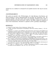
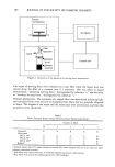
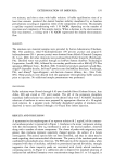
Purchased for the exclusive use of nofirst nolast (unknown)
From: SCC Media Library & Resource Center (library.scconline.org)