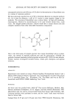
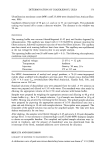
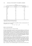
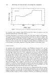
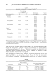
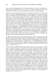
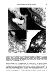
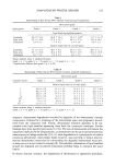
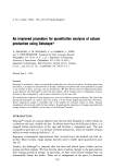
214 JOURNAL OF THE SOCIETY OF COSMETIC CHEMISTS Table III Relationship of Skin Xerosis With Stratum Comeurn Lipid Composition in the Inner and Outer Layers of the Stratum Corneum Skin xerosis grade Grade 1 Grade 2 Lipid species Outer SC Inner SC Outer SC Inner SC Lipid levels (ng lipid/ng protein) Ceramides 65.8 + 19.3 62.5 --+ 21.0 49.8 --- 23.0 64.5 --- 41.0 Fatty acids 53.0 - 30.6 42.5 - 22.9 64.4 -+ 51.7 39.3 +- 20.2 Cholesterol 6.4 --+ 5.0 5.2 --- 4.1 10.0 + 5.0 6.0 --+ 5.0 Relative lipid levels (% of total lipids) Ceramides 52.6 -+ 11.9 56.8 -+ 12.0 45.4 -+ 15.1 58.6 -+ 11.8' Fatty acids 42.4 + 11.9 38.6 -+ 12.6 45.5 -+ 21.6 35.9 +- 14.4 Cholesterol 5.1 -+ 2.5 4.7 -+ 3.1 9.0 + 7.0 5.6 + 3.4 Skin xerosis grade Grade 3 Grade 4 Lipid species Outer SC Inner SC Outer SC Inner SC Lipid levels (ng lipid/ng protein) Ceramides 49.6 + 38.1 47.6 + 22.8 41.7 --- 11.7 47.7 - 35.0 Fatty acids 92.8 - 70.4 53.6 - 38.6 84.2 - 49.8 48.5 -+ 28.8 Cholesterol 7.2 -+ 4.1 6.3 - 7.6 9.5 -+ 4.3 3.5 + 2.5 Relative lipid levels (% of total lipids) Ceramides 34.7 + 18.5 44.7 + 20.8* 35.2 -+ 14.1 45.5 + 10.6' Fatty acids 59.3 + 18.5 46.4 -+ 18.7 57.7 -+ 13.8 49.9 +- 9.4 Cholesterol 6.1 -+ 6.1 6.0 + 4.4 7.1 -+ 1.9 4.6 -+ 2.7' Values represent mean - standard deviation. Inner = tapes 2-8 Outer = tape 1. Grade 1, n = 8 grade 2, n = 8 grade 3, n = 12 grade 4, n = 12. * Significant differences between inner and outer stratum corneum (p 0.05). resulting in intact desmosomes in the surface layers, shown by electron microscopy and analysis of dsg 1 levels. The defective desmosome catabolism is probably the cause of the accumulation of corneocyte clumps on the surface of the skin to produce the scaling condition. Similar findings of desmosomal retention have been demonstrated in other skin scaling disorders, either pathological or experimentally induced (24,40-42). In- terestingly, dsgl appears to be the major adhesive desmosomal protein in the outer layers of the stratum corneum, since the other desmosomal cadherins, the desmocollins, appear to be degraded in the lower layers of the stratum corneum (43). As with the desmosomes, the stratum corneum lipid lamellae appeared to undergo a process of "degradation" or disruption at the surface layers of the cornified layer in normal skin the classical intercellular multiple-lipid bilayers present just below the surface layers appeared to disappear in the surface layers of the stratum corneum. Our findings are supported by infrared spectroscopic methods that found more fluid and

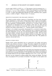
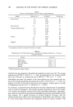
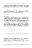
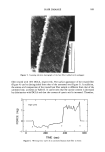
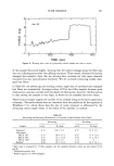
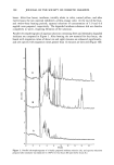
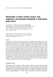
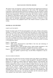
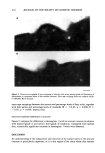
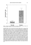
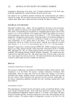
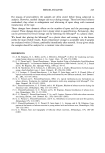
SOAP-INDUCED WINTER XEROSIS 215 6 5 _ o Normal Xerosis Figure 5. Histogram showing the increased levels of desmoglein 1 in stratum corneum of subjects with severe xerosis (grade 4) compared with normal stratum corneum (grade 1). disordered lipids in the outer layers of the stratum corneum (44). In contrast, lipid bilayers have been shown to persist within the intercellular spaces of epidermal cyst desquamated corneocytes (45) however, the desquamating edges of the corneocytes were not examined in that study. The xerotic stratum corneum also appeared to have surface perturbations in lipid structure the normal membrane structure of the lipid lamellae was replaced by a more disorganized region of lipid, suggesting a collapse of the order of the bilayers. Tape stripping is unlikely to cause this effect because it was not seen in tape strippings from normal stratum corneum. Furthermore, it is unlikely that the changes in lipid structure in the outer layers of the stratum corneum were due to residual petrolatum from previously applied moisturizers (46), since the changes in lipid struc- ture were consistent in every subject and the moisturizers used by subjects prior to this study did not contain petrolatum. However, it was possible that the changes in lipid structure were due to superficial extraction of stratum corneum lipids by the cleansing regime, and this is probably the reason for the lower than expected levels of cholesterol in total stratum corneum (47). Imokawa et al. (28) clearly demonstrated the loss of lipids from stratum corneum following surfactant insult. Despite this, the levels of cholesterol in the superficial layers of the stratum corneum are similar to those reported by others (48).
Purchased for the exclusive use of nofirst nolast (unknown)
From: SCC Media Library & Resource Center (library.scconline.org)