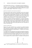
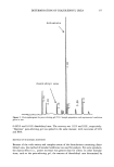
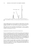
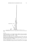
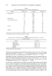
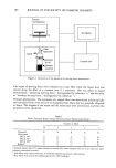
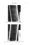
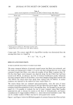
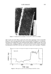
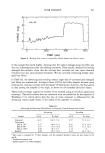
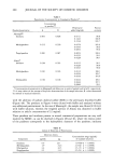
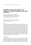
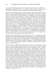
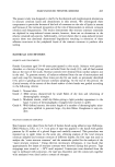
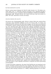
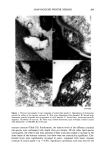
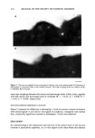
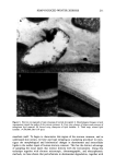
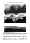
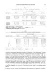
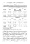
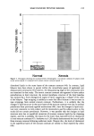
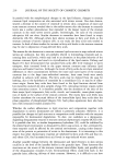
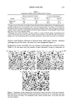
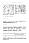
208 JOURNAL OF THE SOCIETY OF COSMETIC CHEMISTS coefficients between each lipid component and the xerosis grades were determined using the Spearman correlation coefficient. RESULTS STRATUM CORNEUM LIPID AND DESMOSOME STRUCTURE IN NORMAL AND XEROTIC SKIN Sequential tape stripping of normal skin revealed morphological changes for stratum corneum lipids and desmosomes in the inner and outer layers of the stratum comeurn. In lower layers (third tape strip down) intact electron-dense desmosomal structures were seen (Figure 1D) in direct contact with the intercellular lipid lamellae. The desmosomes appeared to undergo degradation and a reduction in number in the upper layers of the stratum corneum. During their degradation, desmosomes showed digestion of their internal elements with vacuolation of their structures (Figure 1C) before detaching from the corneocyte envelopes. Desmosomal remnants often appear to be surrounded by intercellular lipids (Figure lB) before their total degradation (Figure 1A). The deeper tissue regions of winter xerotic stratum corneum resembled normal tissue. However, in contrast to normal skin, desmosomes persisted to the surface layer of the stratum corneum (Figure 2). The definition of a normal desmosome in these studies refers to desmosomes with electron-dense internal structures. During the tape-stripping proce- dure, desmosomes may be detached from adjacent cells, as in Figure 1D. In the lower layers of normal stratum corneum, lipids were present as multiple lamellae bilayer structures between the corneocytes (Figure 3C). However, toward the surface layers of the stratum corneum, the bilayer structures were no longer present, and appeared to have been degraded and replaced with a more amorphous-like structure (Figure 3A,B). In severe xerosis (grade 4 Figure 4), normal intercellular lipid structures were found in the lower layers of the stratum corneum (Figure 4C). However, in the peripheral layers of stratum corneum from subjects with severe xerosis, the normal lipid bilayer structure was replaced by large amounts of disorganized intercellular lipids (Figure 4A,B). STRATUM CORNEUM LIPID ANALYSIS An initial analysis of stratum corneum lipid composition from normal and xerotic skin was performed on pooled corneocytes from all of the tape strippings. Compared with normal skin (grade 1), statistically significant decreases in the mass levels of ceramide were seen in xerosis grades 3 and 4 (p 0.05), but not with grade 2 (Table I). Furthermore, the relative levels of the different ceramide species were unchanged (Table II). As changes in lipid structure were apparent in the outer layers of the stratum corneum in winter xerosis in the electron microscopy studies, we analyzed stratum comeurn lipids by depth, comparing the outer stratum corneum (first tape stripping) with the combi- nation of lipids in the remaining tape strippings (inner stratum corneum). No signif- icant difference was detected in the relative levels of ceramides in the outer and inner layers of stratum corneum in normal skin however, significant decreases in the relative levels of ceramide were seen in all grades of xerosis in the outer, compared with inner,

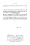
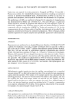
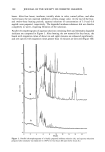
SOAP-INDUCED WINTER XEROSIS 209 A D Figure 1. Electron micrographs of tape strippings of normal skin (grade 1). Degradation of desmosomes toward the surface of the stratum corneum: A. First strip desmosome fully degraded. B. Second strip desmosome partially degraded and encapsulated by lipid lamellae. C. Second strip desmosome partially degraded, vacuolation of structure. D. Third strip normal desmosome lipid envelopes in direct contact with desmosome. (x200,000 Bar 0.05 •m). stratum corneum (Table III). Furthermore, the relative levels of the different ceramide sub-species were unchanged with depth (data not shown). Of the other lipid species investigated, the relative and mass amounts of fatty acids also tended to increase in the outer layers of the stratum corneum, but these were not statistically significant. Cho- lesterol levels were significantly increased in outer, compared with inner, stratum corneum of xerosis grade 4 (p 0.05). Significant correlations were also seen for the
Purchased for the exclusive use of nofirst nolast (unknown)
From: SCC Media Library & Resource Center (library.scconline.org)