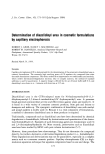
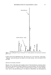
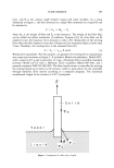
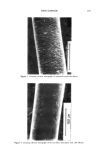
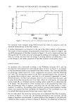
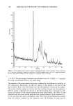
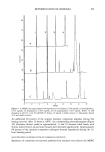
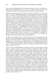
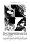
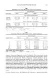
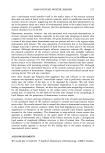
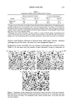
SOAP-INDUCED WINTER XEROSIS 211 Figure 3. Electron micrographs of tape strippings of normal skin (grade 1). Morphological changes in lipid organization toward the surface of the stratum corneum: A. First strip absence of bilayers and presence of amorphous lipid material. B. Second strip disruption of lipid lamellae. C. Third strip normal lipid lamellae. (x200,000 Bar 0.05 manifests itself. To begin to characterize this region of the stratum corneum, and to understand skin xerosis, we have used tape stripping as a sampling procedure to inves- tigate the morphological and biochemical changes in desmosomes and intercellular lipids in the surface layers of human stratum corneum. This has the distinct advantage of sampling the tissue layers that interact directly with the environment. Using this technique together with electron microscopic, chromatographic, and electrophoretic methods, we have shown that perturbations in desmosomal degradation, together with

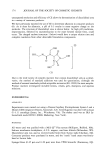
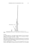
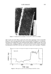
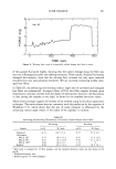
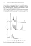
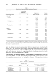
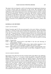
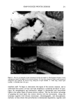
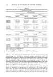
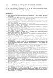
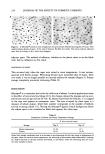
212 JOURNAL OF THE SOCIETY OF COSMETIC CHEMISTS Figure 4. Electron micrograph of tape strippings of subjects with severe xerosis (grade 4). Aberration in lipid organization toward surface of stratum corneum: A. First strip disorganized lipid lamellae. B. Second strip disorganized lipid lamellae. C. Third strip normal lipid lamellae. (x200,000 Bar 0.05 lipid lamellae structure and composition, occur at the stratum corneum surface in soap-induced winter xerosis. In normal skin, desmosomes were found close to the surface, demonstrating persistence of desmosomes throughout most of the stratum corneum. However, degradation of these desmosomes occurred in the most superficial layers of the stratum corneum. In this
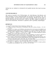
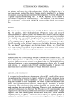
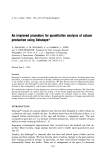
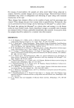
Purchased for the exclusive use of nofirst nolast (unknown)
From: SCC Media Library & Resource Center (library.scconline.org)