60 JOURNAL OF THE SOCIETY OF COSMETIC CHEMISTS sunblocks. Both types of sunscreens aim to reduce the amount of UV light reaching the skin. Two particular difficulties are encountered when formulating physical sunblocks. First, one must select the most appropriate mineral raw material. Among the marketed mineral ingredients appear materials such as titanium dioxide, zinc oxide, iron oxide, and talc (5). These different sunscreen agents may exhibit different sizes and shapes. They also may undergo various surface treatments, already described for usual make-up cosmetic pigments (6). The second problem lies in the incorporation of the mineral into either an aqueous or an oil phase. For instance, a previous paper mentioned the possibility of an irreversible clumping of the mineral agent when used in a powder form (7). Nevertheless, the formulation must provide a good spreadability of the particles that have been worked into the formula. Ideally, a highly efficient physical sunscreen should provide a satisfactory protection against ultraviolet light, be aesthetically acceptable by appearing transparent when applied to the skin, and offer a silky feel. The purpose of this study was to examine the behavior pattern of ultrafine titanium dioxide crystals incorporated in various sunscreen formulations. Two types of TiO 2 were morphologically and crystallographically char- acterized. Three different sunblocks were realized and compared in electron microscopy. The mineral ingredient/cosmetic vehicle relationship as well as the spatial distribution of the metallic oxide particles onto the skin are of utmost interest for the improvement of physical sunscreens. Thus our investigations will undoubtedly contribute to a better knowledge of the reciprocal effects of the different factors. MATERIALS AND METHODS FORMULATION PRINCIPLES Sunscreen preparation 1. Acicular ultrafine titanium dioxide predispersed in mineral oil/ triglyceride (Tioveil MOTG, Tioxide Chemicals Ltd, UK) is encapsulated in oil droplets (H•liosides ©, Av&ne, a Pierre Fabre cosmetic patent) (8). Those microspheres are trapped in a ramified acrylate polymer base, thus yielding a steric stabilization of oil droplets in the aqueous gel. Consequently, the metallic oxides are hermetically isolated from the surrounding aqueous gel. This photoprotective microdispersion resembles an oil/water formula, but has the advantage to be formulated without any emulsifying surfactant. The TiO2 content in the final product = 6 wt %. The SPF is 6.9. Sunscreen-preparation 2. Prismatic ultrafine titanium dioxide powder (VP Titanium Di- oxide T805, Degussa, Germany) is added to the photoprotective microdispersion de- scribed above (sunscreen preparation 1). The prismatic oxides are confined in the oil microspheres (H•liosides©), thus separated from the acrylate gel. The TiO2 content in the final product = 6 wt %. The SPF is 8.7. Sunscreen preparation 3. The prismatic ultrafine TiO2 mentioned above (sunscreen prep- aration 2) is incorporated in a water/oil emulsion (a Pierre Fabre cosmetic patent). Optimal dispersion of the oxide particles is achieved by thorough grinding procedures within different specific esters and oils, thus breaking up the TiO 2 agglomerates. This total sunblock is formulated only with physical sunscreen agents. The TiO2 content in the finished product = 6 wt %. The SPF is 12.9.

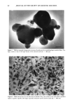
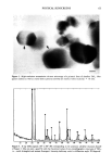
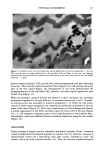
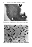
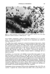
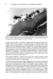
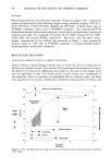
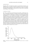
PHYSICAL SUNSCREENS 61 In vivo sun protection factors (SPF). These were determined in accordance with COLIPA method (9) on five subjects for each product using the standard P3 (Bayer, reference C202/101), giving in our case a SPF value of 18.4. INVESTIGATION PROCEDURES Scanning electron microscopy. A droplet containing H61iosides © in an aqueous solution was deposited onto a coverglass and was subsequently vacuum dried for eight hours. Finally the preparation underwent a gold-palladium coating (Hummer-junior, Siemens, Ger- many). The observations were realized with a JEOL JSM 35C (JEOL, Japan) scanning electron microscope (SEM) at 25 kV. Transmission electron microscopy. For the morphological and crystallographical assessments of the raw material, the TiO2 particles were deposited directly on 1000-mesh copper grids covered with a carbon-coated formvar film. The samples were investigated in a JEOL EM 100B (JEOL, Japan) transmission electron microscope (TEM), equipped with a nitrogen-cooled anticontamination device, at 100 kV. The following scheme was used for the preparation of skin surface biopsies: Physical sunscreen products were carefully rubbed for 15 seconds into the test area of the inner forearm of one male volunteer subject. Twenty minutes later, 10 to 15 mm 2 large and cyanoacrylate glue-coated plastic strips were gently pressed to the creamed areas for one minute. The recovered plastic strips including the outer horny layer of the treated skin areas were immediately fixed in a 2% paraformaldehyde-glutaraldehyde solution buffered at pH 7.4 with 0.1 sodium cacodylate. Ultrathin sections were achieved, from the embedded (Epon 812) surface biopsies, by using a MT-2C Sorvall-Porter ultramicrotome equipped with a diamond knife. When necessary, staining of the sections was performed with uranyl acetate and lead citrate. The sections were examined in TEM (JEOL EM 100B) with an accelerating voltage of the electrons of 60 kV. XRD analysis. X-ray diffraction was performed on the raw material (TiO2 prior to formulation) with a Kristalloflex D500 (Siemens, Germany) using the CuK• radiation (kCuK• = 0. 154 nm). Experimental data were compared to reference values of the Joint Committee of Powder Diffraction Standards (JCPDS). RESULTS The so-called pigmentary titanium dioxide particles, which are widely used in paints for their light-scattering properties, measure about 300 nm in diameter (Figure 1). In order to gain optimum benefit, the TiO• crystallites employed for the formulation of physical sunscreens must present much smaller sizes. Although these commercially available ultrafine oxides generally correspond to the required sizes, they may exhibit different morphological and crystallographical characteristics. Figure 2 shows an acicular form of TiO2 crystals, whereas platelet-like habits of an other ultrafine titanium dioxide are visible in Figure 3. The resolving power of the TEM allowed us to visualize intersecting sets of lattice fringes in several nanocrystals of TiO• (Figure 3). TiO2 raw material may also be a mixture of different crystallographic forms of TiO2, e.g., rutile and anatase, brookire being a third but quite rare structure. The crystallographic features were determined by X-ray diffraction. For instance, Figure 4 is the powder diffraction dia-
Purchased for the exclusive use of nofirst nolast (unknown)
From: SCC Media Library & Resource Center (library.scconline.org)