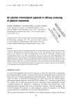
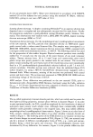
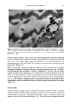
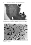
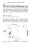
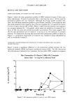
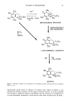
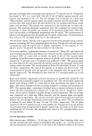
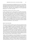
62 JOURNAL OF THE SOCIETY OF COSMETIC CHEMISTS Figure 1. TEM micrograph ofpigmentary titanium dioxide particles resembling large roundish flakes. The high contrast is due to the high density of the mineral (scale bar = 100 nm). Figure 2. TEM observagion of an acicular form of ultrafine TiO 2. Sets of four or five individual crystallites appear to gather together into larger equi-axed structures (curved arrows) (scale bar = 100 nm).

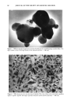
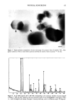
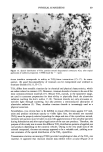
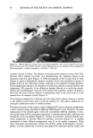
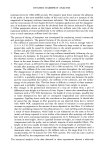
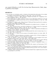
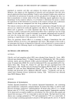
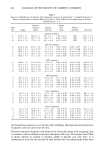
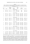
PHYSICAL SUNSCREENS 63 Figure 3. High-resolution transmission electron microscopy of a primatic form of ultrafine TiO 2 Hex- agonal outlines as well as crystal lattice patterns (arrows) are clearly evident (scale bar = lO nm). Figure 4. X-ray diffractogram (10 ø to 80 ø 20) corresponding to the prismatic ultrafine titanium dioxide of Figure 3 One can note a good fit with the theoretical values of two crystallographic structures of TiO 2, i.e., rutile (triangles) and anatase (lozenges). Intensity (arbitrary units) vs diffraction angle 20 (degrees).
Purchased for the exclusive use of nofirst nolast (unknown)
From: SCC Media Library & Resource Center (library.scconline.org)