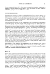
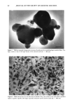
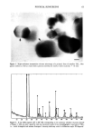
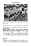
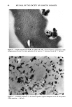
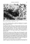
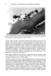
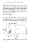
64 JOURNAL OF THE SOCIETY OF COSMETIC CHEMISTS .w Figure 5. In SEM the H•liosides © appear as well-defined individual microspheres (scale bar = 100 •m). gram corresponding to the prismatic form of titanium dioxide observed in Figure 3. These XRD data match with both crystallographic forms of TiO 2 i.e., rutile (JCPDS file number 21-1276) and anatase LICPDS file number 21-1272). The large depth of field of SEM allows us to visualize the three-dimensional aspects of a particular cosmetic vehicle (H•liosides ©) used in sunscreen preparations 1 and 2. The mineral ingredients appear to be trapped in spherical structures, as shown in Figure 5. The spherical bodies (H•liosides ©, in preparations 1 and 2) have various sizes, with an average diameter of 2.58 •xm (standard deviation, cr = 1.05 •xm). Two selected TiO 2 raw materials (acicular and prismatic) were incorporated in those oil droplets (H•lio- sides©). When the acicular form of TiO2 was combined with the photoprotective microdisper- sion in sunscreen preparation 1, the crystallites appeared to be mainly confined to the periphery of the oil droplets. Some of those microspheres could even be observed after topical application to skin (Figure 6). Nevertheless, many areas showed a thin and regular layer of acicular TiO 2 crystallites lining the stratum corneum (Figure 7). When the prismatic TiO 2 powder was combined with the same photoprotective micro- dispersion in sunscreen preparation 2, it was again possible to visualize more or less spherical arrangements. However, in this case the crystals seemed evenly distributed in the oil droplets, thus occupying the whole room offered by the microspheres. This aspect was particularly evident along the laminar cells of hair cuticles (Figure 8). This different behavior of the mineral within the oil droplets (H•liosides ©) might be explained by the hydrophobic character of this prismatic titanium dioxide. After application of the photoprotective microdispersion onto skin, the burst micro-

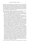
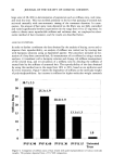
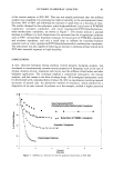
PHYSICAL SUNSCREENS 65 Figure 6. Ultrathin section of a skin biopsy after topical application of sunscreen preparation 1. Acicular TiO 2 crystals seem to arrange preferentially at the periphery of the oil phase. In this case, one spherical microstructure has penetrated the widened intercellular spaces of the outer horny layer of the skin (TEM) (scale bar = l •m). spheres released prismatic TiO 2 crystals that could homogeneously coat the underlying structures. This could be verified around hair fibers (Figure 9) as well as along the horny layer of the skin surface (Figure 10). Enlargements of such areas demonstrated the nonagglomeration of the individual TiO2 particles, even after topical application onto human skin (Figure 11). When the prismatic titanium dioxide was added to a classic emulsion, the crystalline arrangement appeared to be quite different in sunscreen preparations 1 and 2. Actually, no microstructure was noticeable in sunblock preparation 3- In TEM, the tiny mono- crystals or small clusters appeared to be randomly and uniformly distributed in the oily phase of the cream (Figure 12). After topical application we could subsequently observe a similar organization of the TiO2 crystallites upon the horny cells of the skin (Figure 13), i.e., the inorganic component more or less evenly dispersed in the emulsion layer. Nevertheless, some areas exhibited thinner crystalline depositions along the skin surface (Figure 14). DISCUSSION Today titanium is largely used for orthopedic and dental implants. Hence, numerous studies considered the histological responses to titanium (10-13). However, titanium is spontaneously covered by a passivating oxide layer mainly composed of rutile and anatase, which are both titanium dioxides (14). Thus the titanium implant/biological
Purchased for the exclusive use of nofirst nolast (unknown)
From: SCC Media Library & Resource Center (library.scconline.org)