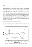
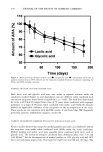
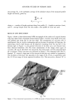
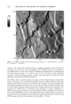
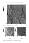
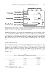
AFM FOR STUDIES OF HUMAN HAIR 201 ness average, Ra, is the arithmetic average of the absolute values of the measured profile height deviations, given by: n •lZ i - Zl i=l Ra = (1) n where, n -- number of height positions along line profile, Z i -- height at position i (nm), -- and Z -- average height (nm). For this study, a line profile of 4 pm was used. RESULTS AND DISCUSSION Figure 1 shows a three-dimensional AFM micrograph of the surface of a typical human scalp hair taken from a European brown-haired subject. Cuticles were found to be very well defined and lying flat on the hair shaft. The arrangement of cuticular cells is multilayered, due to cuticles overlapping and not because of multiple cell layering. An interesting scratch mark feature can be observed traversing from the top left to the bottom right in Figure 1 in the general direction of the fiber axis such features have been observed elsewhere (19). On closer examination of the image, three types of cuticular cell can be identified: the exocuticle, the endocuticle, and the marginal band (A- or u-layer). These structures are indicated in Figure 2. The marginal band (20,21), denoted "A" covered a large part of the exocuticle and was very smooth in appearance. The thickness of this layer was typically in the range of 30-70 nm, in agreement with the 45-50 nm range of values reported elsewhere (22). The exocuticles, denoted "B" in ..-: • [ 26,52 23 nm 1326.12 nm 0 nm . '.." i :'-._ ,,. ß .. ß . . 20•m•......... '. : . , •' , . Ol•m Ol•fn Figure 1. Three-dimensional representation of an AFM micrograph of human European brown scalp hair.

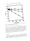
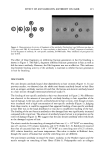
202 JOURNAL OF THE SOCIETY OF COSMETIC CHEMISTS ß -90.0 ø 90.0 ø 20 prn .... ,' ( 10 prn A 0pm , ,: ::-,, 0 prn B B A B B 10 pm 20 pm Figure 2. An AFM micrograph of human European brown scalp hair. A = marginal band (A- or c•-layer), B = exocuticle, C = endocuticle. Figure 2, were much more rough, showing a woodgrain appearance in the direction of the fiber axis. Exocuticles were very jagged at their broken distal ends, in agreement with SEM studies. In some cases, where the breakage was fragmented, a rougher deposit of endocuticular material, "C," could be seen on the top surface of the underlying exocuticle. Similar breakage patterns, together with postulated mechanisms of their formation, have been discussed elsewhere (3). The AFM is able to provide quantitative data as well as high-resolution visual infor- mation. In Figure 3, two cross-sectional profiles show the surface relief of an exocuticle (upper) and an endocuticle (lower). As expected, the exocuticle was much smoother than the exposed underlying endocuticle (Ra: 9 nm cf. 30 nm). The use of surface profilometry to record surface roughness along the fiber axis of human hairs has been reported in the literature (23). While there are obviously some similarities in surface profilometry to AFM, the macroscopic technique is much less sensitive. When operating on a submicron scale, height information can be grossly affected by the very large radius of curvature of the tip (2.5 Fm). Such artefacts are not observed in profile measurements obtained using AFM, because the radius of curvature is much smaller (50 nm).
Purchased for the exclusive use of nofirst nolast (unknown)
From: SCC Media Library & Resource Center (library.scconline.org)