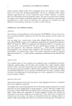
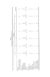
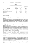
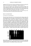
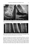
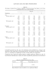
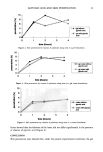
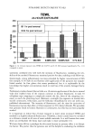
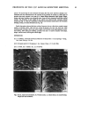
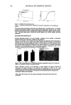
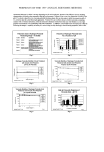
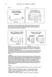
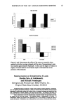
DAMAGED COMPONENTS OF PERMED HAIR 21 Table IV Changes in the Half-Cystine Contents of Each Fraction Toward the Tip End • Length Hair type (cm) Matrix Microfibril Residue + cuticle No treatment 0-10 25.0 + 4.5 b 9.2 _+ 3.2 10-20 23.6 -+ 2.9 9.2 -+ 2.9 20-30 22.4 + 3.1 10.0 + 3.0 30•40 22.4 _+ 3.3 9.7 + 1.8 Type I 0-10 25.4 _+ 5.5 10.8 _+ 2.3 10-20 22.4 _+ 5.5 7.2 + 4.5 20-30 21.2 _+ 6.0 6.6 + 5.1 30-40 20.1 + 2.5 6.0 + 6.2 Type II 0-10 20.2 _+ 4.9 11.9 + 3.7 10-20 19.8 + 4.1 11.6 + 2.7 20-30 18.9 + 3.9 11.4 _+ 4.2 30-40 18.9 -+ 3.6 11.5 + 5.7 16.4 _+ 3.3 15.6 + 4.2 15.2 +_ 5.8 15.1 _+ 4.9 15.8 + 3.8 15.5 _+ 2.9 12.7 + 5.3 10.0 + 4.7 19.5 -+ 4.1 16.8 ñ 4.4 15.8 + 6.7 10.4 + 4.6 All values are the mean of three experiments. Refer to Figure 1 for experimental conditions. • Estimated as cysteic acid and expressed as residues/100 amino acids. b+SD. reported that the disulfide bond reformation with peroxide following perming is not complete and that the cysteic acid content in the perreed hair is higher than that in untreated hair. Our results showed similar concerns with the Type I hair, but the Type II hair showed an interesting result: although the residue (including the cuticle) was also oxidized just as in Type I, the cortical protein was not oxidized like the untreated hair. In other words, in the split-end hair, not only the cuticle but also the cortical proteins were oxidized. Table V Changes in the Cysteic Acid Contents of Each Fraction Toward the Tip End • Length Hair type (cm) Matrix Microfibril Residue + cuticle No treatment 0-10 3.7 + 0.33 2.5 + 0.1 10-20 4.5 + 0.4 3.0 _+ 0.9 20-30 5.3 + 1.1 3.3 + 0.6 30-40 7.2 + 0.9 4.6 + 0.5 Type I 0-10 3.2 -+ 1.2 2.6 _+ 1.1 10-20 8.4 + 2.1 6.7 + 1.3 20-30 16.3 + 1.3 12.3 + 0.8 30-40 17.5 _+ 0.9 14.5 + 2.4 Type II 0-10 1.6 + 1.5 0.2 + 0.2 10-20 2.7 _+ 0.7 1.4 + 0.8 20-30 4.5 + 0.6 2.4 _+ 0.3 30-40 6.3 + 2.7 2.8 + 1.1 4.2 + 0.3 4.3 + 0.6 4.9 + 0.6 6.3 -+ 1.0 4.3 + 7.5 + 11.7 + 13.1 + 4.7 + 8.8 _+ 12.4 _+ 13.0 + 0.8 1.2 0.7 3.4 2.4 1.3 4.4 2.9 All values are the mean of three experiments. Refer to Figure 1 for experimental conditions. • Expressed as residues/1000 amino acids. •+SD.

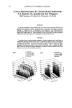
22 JOURNAL OF COSMETIC SCIENCE CONCLUSION We have developed a novel method for quantifying hair components. This method permits the detailed analysis of components damaged due to perming. In summary, we have found a significant decrease in microfibril protein and an increase in high- molecular-weight protein on the tip end of permed hair. We suggest that the "intact" microfibril protein turns into high-molecular-weight protein due to perming. Moreover, we have found that the cuticle was not so damaged, though the cortical proteins were oxidized in the split-end hair. Using this method, additional work is currently being undertaken in order to ascertain the correlation between the morphological changes and the degree of damage. REFERENCES (1) C. M. Pande and J. Jachowicz, Hair photodamage--Measurement and prevention,J. Soc. Cosmet. Chem., 44, 109-122 (1993). (2) S. E. Kelly and V. N. E. Robbinson, The effect of grooming on the cuticle, J. Soc. Cosmet. Chem., 33, 203-215 (1982). (3) J. A. Swift and A. C. Brown, The critical determination of the fine changes in the surface architecture of human hair due to cosmetic treatment, J. Soc. Cosmet. Chem., 23, 695-702 (1972). (4) C. R. Robbins, Chemical and Physical Behavior of Human Hair, 3rd ed. (Springer-Verlag, New York, 1994), pp. 211-226. (5) S. H. Bong and H. Zahn, Contributions to the chemistry of human hair. II. Lipid chemical aspects of permanently waved hair, Int, J. Cosmet. Sci., 11, 167-174 (1989). (6) J. Chao, A. E. Newsom, I. M. Wainwright, and R. A. Mathews, Comparisons of the effects of some reactive chemicals on the proteins of whole hair, cuticle and cortex,J. Soc. Cosmet. Chem., 30, 401-413 (1979). (7) R. C. Marshall and J. M. Gillespie, Comparison of samples of human hair by two dimensional elec- trophoresis,J. Forensic. Sci. Soc., 22, 377-388 (1982). (8) C. Nappe and M. Kermici, Electrophoretic analysis of alkylated proteins of human hair from various ethnic groups,J. Soc. Cosmet. Chem., 40, 91-99 (1989). (9) J. M. Gillespie, "The Structure Proteins of Hair: Isolation, Characterization, and Regulation of Bio- synthesis" in Biochemistry and Physiology of the Skin, L. A. Goldsmith, Ed. (Oxford University Press, London, 1983), Vol. 1, pp. 475-510. (10) H. Shiigger and G. U. Jagow, Tricine-sodium dodecyl sulfate-polyacrylamide gel electrophoresis for the separation of proteins in the range from 1 to 100 kDa, Anal. Biochem., 166, 368-379 (1987). (11) J. Adamski, B. Husen, H. H. Thole, U. G. Stewart, and P. W. Jungblut, Linkage of 1713-oestradiol dehydrogenase to actin by e-(y-glutamyl)-lysine in porcine endometrial cells, Biochem. J., 296, 797- 802 (1993). (12) P. W. Wertz and D. T. Downing, Integral lipids of human hair, Lipids, 23, 878-881 (1988). (13) W. Diinges, 4-Bromomethyl-7-methoxycoumarine as a new fluorescence label for fatty acids, Anal. Chem., 49, 442-445 (1977). (14) J. A. Swift, Chemical composition of various morphological components isolated from human hair cuticle, Cosmet. Toiletr., 91, 46-48 (1976). (15) J. A. Swift and B. Bews, The chemistry of human hair cuticle. I. A new method for the physical isolation of cuticle. J. Soc. Cosmet. Chem., 25, 13-22 (1974). (16) A. Nakamura, R. Kon, and K. Takeuchi,Japanese Patent (submitted). (17) H. Zahn, Wool chemistry and processing, Abstracts of Proc. 9th Int. Wool Textile Res. Conf (Biella), 1995, pp. 1-16. (18) H. Zahn, Wool is not keratin only, Abstracts ofProc. 6th Int. Wool Textile Res. Conf (Pretoria), 1980, pp. 1-45. (19) N. Yorimoto and S. Naito, Physical and chemical properties of integral lipids in hair cell membrane complex, Proc. ISF '94 (Yokohama), preprint, 1994, p. 215.
Purchased for the exclusive use of nofirst nolast (unknown)
From: SCC Media Library & Resource Center (library.scconline.org)